Jairo Alfonso Mendoza-Roldan, Mariaelisa Carbonara, Viviane Noll Louzada-Flores, Mario H Alves, Nicola Pugliese, Nicola Decaro, Annamaria Uva, Floriana Gernone, Maria Alfonsa Cavalera, Andrea Zatelli, Domenico Otranto
{"title":"户外猫的幼利什曼原虫和沙罗利什曼原虫及猫源性外周血单个核细胞感染报告。","authors":"Jairo Alfonso Mendoza-Roldan, Mariaelisa Carbonara, Viviane Noll Louzada-Flores, Mario H Alves, Nicola Pugliese, Nicola Decaro, Annamaria Uva, Floriana Gernone, Maria Alfonsa Cavalera, Andrea Zatelli, Domenico Otranto","doi":"10.1186/s13071-025-06983-w","DOIUrl":null,"url":null,"abstract":"<p><strong>Background: </strong>Feline leishmaniosis (FeL) is mainly caused by Leishmania infantum in the Mediterranean Basin. In Italy, in the same epidemiological context where canine leishmaniosis (CanL) is hyperendemic, a nonpathogenic species, Leishmania tarentolae, may also occur in sympatry, infecting reptiles, dogs, and humans. Thus, this study aimed to assess L. tarentolae infection in outdoor cats along with its co-occurrence with L. infantum and to evaluate risk factors. In addition, the persistence of L. tarentolae in feline-derived peripheral blood mononuclear cells (PBMCs) was herein evaluated in vitro.</p><p><strong>Methods: </strong>Outdoor colony or stray cats were screened for Leishmania spp. by immunofluorescence antibody test (IFAT) using promastigotes of both L. infantum and L. tarentolae. Whole blood and buffy coat were tested by a real-time polymerase chain reaction (qPCR) and duplex real-time PCR (dqPCR), and positive samples sequenced following an ITS1 conventional PCR (cPCR). Feline-derived PMBCs were subsequently infected with promastigotes of L. tarentolae to assess the persistence of amastigotes. Viral infections caused by feline immunodeficiency virus (FIV) and feline leukemia virus (FeLV) were molecularly addressed in all enrolled cats. Statistical analysis was performed to evaluate the possible association between Leishmania spp. infection and FIV/FeLV infection by using a multivariate logistic regression model following an initial LASSO-penalized logistic regression.</p><p><strong>Results: </strong>Overall, 42 out of 194 cats (21.6%) were serologically or molecularly positive for Leishmania spp. In particular, 26 (13.4%) cats were seropositive for L. infantum and/or L. tarentolae by IFAT, with 16 (8.2%) animals positive for both species. Molecularly, 14 out of 194 cats (7.2%) were positive for L. infantum by qPCR, whereas five (2.6%) were positive for L. tarentolae by dqPCR. Cat PBMCs were successfully infected with L. tarentolae, and the infection persisted for at least 72 h. Overall, 38 out of the 194 screened cats (19.6%) were infected by FIV and/or FeLV, of which 12 were serologically or molecularly positive for Leishmania spp., with one cat positive for L. tarentolae DNA, and five for L. infantum DNA. Multivariate screening identified municipality (OR 2.206; P = 0.031; 95% CI 1.077-4.516) as a risk factor for Leishmania spp. infection, while the association between Leishmania spp. and FIV infection was not significant (OR 2.359; P = 0.08, 95% CI 0.901-6.179).</p><p><strong>Conclusions: </strong>Colony or stray cats were herein found for the first time infected by L. tarentolae, in areas where L. infantum is endemic. Cross-reactivity using IFAT test may pose a diagnostic hindrance also in FeL. The infection with this saurian-associated Leishmania in cats was further confirmed through the persistence of this Leishmania in cat PBMCs. Further studies are needed to fully unravel the complex interactions between both species of Leishmania and the implication of the sympatric occurrence of both species in the diagnosis and control of leishmaniosis.</p>","PeriodicalId":19793,"journal":{"name":"Parasites & Vectors","volume":"18 1","pages":"361"},"PeriodicalIF":3.5000,"publicationDate":"2025-08-26","publicationTypes":"Journal Article","fieldsOfStudy":null,"isOpenAccess":false,"openAccessPdf":"https://www.ncbi.nlm.nih.gov/pmc/articles/PMC12382112/pdf/","citationCount":"0","resultStr":"{\"title\":\"Leishmania (Leishmania) infantum and Leishmania (Sauroleishmania) tarentolae in outdoor cats and report of infection in feline-derived peripheral blood mononuclear cells.\",\"authors\":\"Jairo Alfonso Mendoza-Roldan, Mariaelisa Carbonara, Viviane Noll Louzada-Flores, Mario H Alves, Nicola Pugliese, Nicola Decaro, Annamaria Uva, Floriana Gernone, Maria Alfonsa Cavalera, Andrea Zatelli, Domenico Otranto\",\"doi\":\"10.1186/s13071-025-06983-w\",\"DOIUrl\":null,\"url\":null,\"abstract\":\"<p><strong>Background: </strong>Feline leishmaniosis (FeL) is mainly caused by Leishmania infantum in the Mediterranean Basin. In Italy, in the same epidemiological context where canine leishmaniosis (CanL) is hyperendemic, a nonpathogenic species, Leishmania tarentolae, may also occur in sympatry, infecting reptiles, dogs, and humans. Thus, this study aimed to assess L. tarentolae infection in outdoor cats along with its co-occurrence with L. infantum and to evaluate risk factors. In addition, the persistence of L. tarentolae in feline-derived peripheral blood mononuclear cells (PBMCs) was herein evaluated in vitro.</p><p><strong>Methods: </strong>Outdoor colony or stray cats were screened for Leishmania spp. by immunofluorescence antibody test (IFAT) using promastigotes of both L. infantum and L. tarentolae. Whole blood and buffy coat were tested by a real-time polymerase chain reaction (qPCR) and duplex real-time PCR (dqPCR), and positive samples sequenced following an ITS1 conventional PCR (cPCR). Feline-derived PMBCs were subsequently infected with promastigotes of L. tarentolae to assess the persistence of amastigotes. Viral infections caused by feline immunodeficiency virus (FIV) and feline leukemia virus (FeLV) were molecularly addressed in all enrolled cats. Statistical analysis was performed to evaluate the possible association between Leishmania spp. infection and FIV/FeLV infection by using a multivariate logistic regression model following an initial LASSO-penalized logistic regression.</p><p><strong>Results: </strong>Overall, 42 out of 194 cats (21.6%) were serologically or molecularly positive for Leishmania spp. In particular, 26 (13.4%) cats were seropositive for L. infantum and/or L. tarentolae by IFAT, with 16 (8.2%) animals positive for both species. Molecularly, 14 out of 194 cats (7.2%) were positive for L. infantum by qPCR, whereas five (2.6%) were positive for L. tarentolae by dqPCR. Cat PBMCs were successfully infected with L. tarentolae, and the infection persisted for at least 72 h. Overall, 38 out of the 194 screened cats (19.6%) were infected by FIV and/or FeLV, of which 12 were serologically or molecularly positive for Leishmania spp., with one cat positive for L. tarentolae DNA, and five for L. infantum DNA. Multivariate screening identified municipality (OR 2.206; P = 0.031; 95% CI 1.077-4.516) as a risk factor for Leishmania spp. infection, while the association between Leishmania spp. and FIV infection was not significant (OR 2.359; P = 0.08, 95% CI 0.901-6.179).</p><p><strong>Conclusions: </strong>Colony or stray cats were herein found for the first time infected by L. tarentolae, in areas where L. infantum is endemic. Cross-reactivity using IFAT test may pose a diagnostic hindrance also in FeL. The infection with this saurian-associated Leishmania in cats was further confirmed through the persistence of this Leishmania in cat PBMCs. Further studies are needed to fully unravel the complex interactions between both species of Leishmania and the implication of the sympatric occurrence of both species in the diagnosis and control of leishmaniosis.</p>\",\"PeriodicalId\":19793,\"journal\":{\"name\":\"Parasites & Vectors\",\"volume\":\"18 1\",\"pages\":\"361\"},\"PeriodicalIF\":3.5000,\"publicationDate\":\"2025-08-26\",\"publicationTypes\":\"Journal Article\",\"fieldsOfStudy\":null,\"isOpenAccess\":false,\"openAccessPdf\":\"https://www.ncbi.nlm.nih.gov/pmc/articles/PMC12382112/pdf/\",\"citationCount\":\"0\",\"resultStr\":null,\"platform\":\"Semanticscholar\",\"paperid\":null,\"PeriodicalName\":\"Parasites & Vectors\",\"FirstCategoryId\":\"3\",\"ListUrlMain\":\"https://doi.org/10.1186/s13071-025-06983-w\",\"RegionNum\":2,\"RegionCategory\":\"医学\",\"ArticlePicture\":[],\"TitleCN\":null,\"AbstractTextCN\":null,\"PMCID\":null,\"EPubDate\":\"\",\"PubModel\":\"\",\"JCR\":\"Q1\",\"JCRName\":\"PARASITOLOGY\",\"Score\":null,\"Total\":0}","platform":"Semanticscholar","paperid":null,"PeriodicalName":"Parasites & Vectors","FirstCategoryId":"3","ListUrlMain":"https://doi.org/10.1186/s13071-025-06983-w","RegionNum":2,"RegionCategory":"医学","ArticlePicture":[],"TitleCN":null,"AbstractTextCN":null,"PMCID":null,"EPubDate":"","PubModel":"","JCR":"Q1","JCRName":"PARASITOLOGY","Score":null,"Total":0}
引用次数: 0
摘要
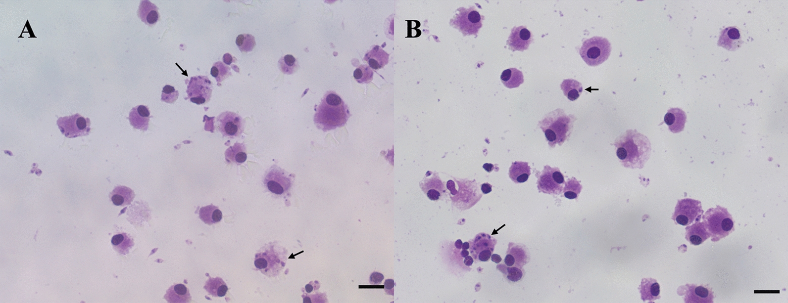
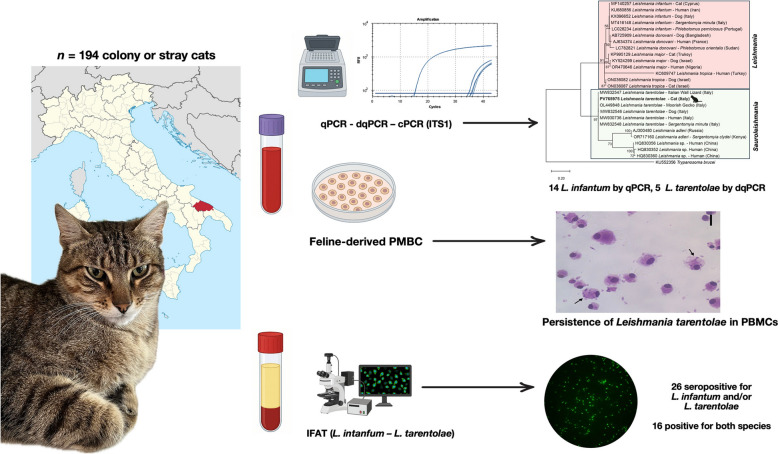
背景:猫利什曼病(FeL)主要由地中海盆地的幼利什曼原虫引起。在意大利,在犬利什曼病(CanL)高地方性流行的相同流行病学背景下,一种非致病性利什曼绦虫(Leishmania tarentolae)也可能在同系动物中发生,感染爬行动物、狗和人类。因此,本研究旨在评估户外猫的猪链乳杆菌感染及其与婴儿乳杆菌的共存情况,并评估其危险因素。此外,本文还对猫源性外周血单个核细胞(PBMCs)中链乳杆菌的持久性进行了体外评估。方法:采用免疫荧光抗体试验(IFAT)对室外集落或流浪猫进行利什曼原虫检测,检测对象为婴儿利什曼原虫和荚膜利什曼原虫。采用实时聚合酶链反应(real-time polymerase chain reaction, qPCR)和双相实时PCR (duplex real-time PCR, dqPCR)检测全血和灰白色被毛,阳性样品采用ITS1常规PCR (conventional PCR, cPCR)测序。随后,猫源性pmbc感染了猪链乳杆菌的原鞭毛菌,以评估无鞭毛菌的持久性。猫免疫缺陷病毒(FIV)和猫白血病病毒(FeLV)引起的病毒感染在所有入选的猫中进行了分子定位。在初始lasso惩罚逻辑回归之后,采用多元逻辑回归模型进行统计分析,以评估利什曼原虫感染与FIV/FeLV感染之间可能的关联。结果:194只猫中有42只(21.6%)对利什曼原虫血清或分子检测呈阳性,其中26只(13.4%)对婴儿乳杆菌和/或链托菌血清检测呈阳性,其中16只(8.2%)对两种细菌均呈阳性。从分子结构上看,194只猫中有14只(7.2%)对婴儿乳杆菌呈qPCR阳性,而5只(2.6%)对猪链乳杆菌呈dqPCR阳性。结果显示,194只猫中有38只(19.6%)感染了FIV和/或FeLV,其中12只在血清学或分子学上呈利什曼原虫阳性,1只猫呈链状乳杆菌DNA阳性,5只猫呈婴儿链状乳杆菌DNA阳性。多因素筛查发现,城市(OR 2.206; P = 0.031; 95% CI 1.077-4.516)是利什曼原虫感染的危险因素,而利什曼原虫和FIV感染之间的相关性不显著(OR 2.359; P = 0.08, 95% CI 0.901-6.179)。结论:在婴幼儿乳杆菌流行地区,首次发现有群猫或流浪猫感染。使用IFAT试验的交叉反应性也可能对FeL造成诊断障碍。通过在猫pbmc中持续存在这种利什曼原虫,进一步证实了猫感染这种与蜥蜴相关的利什曼原虫。需要进一步的研究来充分揭示两种利什曼原虫之间复杂的相互作用,以及两种原虫同地发生在利什曼原虫病的诊断和控制中的意义。
Leishmania (Leishmania) infantum and Leishmania (Sauroleishmania) tarentolae in outdoor cats and report of infection in feline-derived peripheral blood mononuclear cells.
Background: Feline leishmaniosis (FeL) is mainly caused by Leishmania infantum in the Mediterranean Basin. In Italy, in the same epidemiological context where canine leishmaniosis (CanL) is hyperendemic, a nonpathogenic species, Leishmania tarentolae, may also occur in sympatry, infecting reptiles, dogs, and humans. Thus, this study aimed to assess L. tarentolae infection in outdoor cats along with its co-occurrence with L. infantum and to evaluate risk factors. In addition, the persistence of L. tarentolae in feline-derived peripheral blood mononuclear cells (PBMCs) was herein evaluated in vitro.
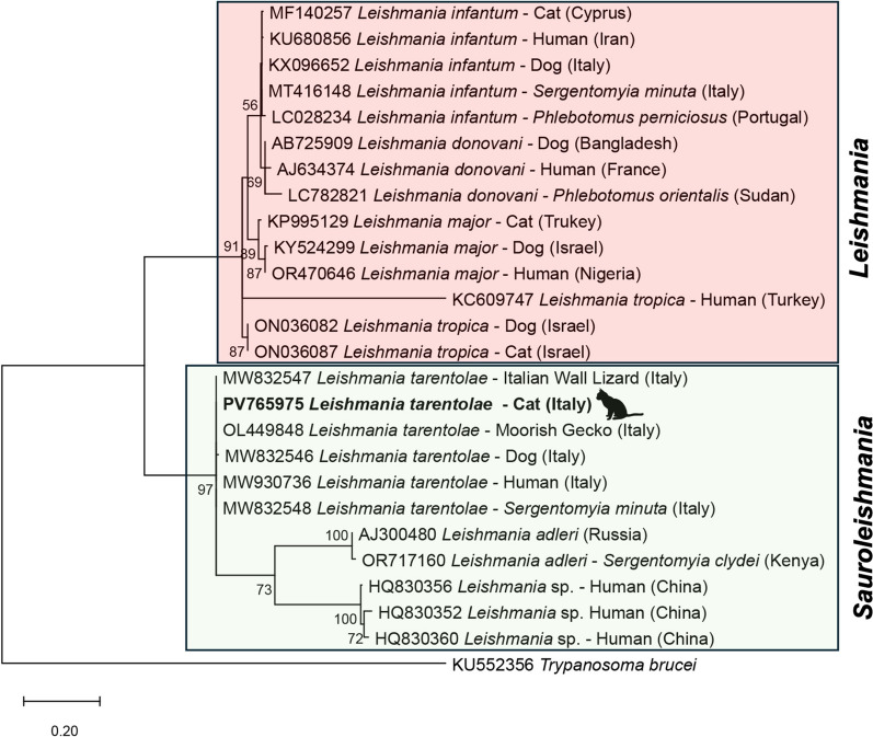
Methods: Outdoor colony or stray cats were screened for Leishmania spp. by immunofluorescence antibody test (IFAT) using promastigotes of both L. infantum and L. tarentolae. Whole blood and buffy coat were tested by a real-time polymerase chain reaction (qPCR) and duplex real-time PCR (dqPCR), and positive samples sequenced following an ITS1 conventional PCR (cPCR). Feline-derived PMBCs were subsequently infected with promastigotes of L. tarentolae to assess the persistence of amastigotes. Viral infections caused by feline immunodeficiency virus (FIV) and feline leukemia virus (FeLV) were molecularly addressed in all enrolled cats. Statistical analysis was performed to evaluate the possible association between Leishmania spp. infection and FIV/FeLV infection by using a multivariate logistic regression model following an initial LASSO-penalized logistic regression.
Results: Overall, 42 out of 194 cats (21.6%) were serologically or molecularly positive for Leishmania spp. In particular, 26 (13.4%) cats were seropositive for L. infantum and/or L. tarentolae by IFAT, with 16 (8.2%) animals positive for both species. Molecularly, 14 out of 194 cats (7.2%) were positive for L. infantum by qPCR, whereas five (2.6%) were positive for L. tarentolae by dqPCR. Cat PBMCs were successfully infected with L. tarentolae, and the infection persisted for at least 72 h. Overall, 38 out of the 194 screened cats (19.6%) were infected by FIV and/or FeLV, of which 12 were serologically or molecularly positive for Leishmania spp., with one cat positive for L. tarentolae DNA, and five for L. infantum DNA. Multivariate screening identified municipality (OR 2.206; P = 0.031; 95% CI 1.077-4.516) as a risk factor for Leishmania spp. infection, while the association between Leishmania spp. and FIV infection was not significant (OR 2.359; P = 0.08, 95% CI 0.901-6.179).
Conclusions: Colony or stray cats were herein found for the first time infected by L. tarentolae, in areas where L. infantum is endemic. Cross-reactivity using IFAT test may pose a diagnostic hindrance also in FeL. The infection with this saurian-associated Leishmania in cats was further confirmed through the persistence of this Leishmania in cat PBMCs. Further studies are needed to fully unravel the complex interactions between both species of Leishmania and the implication of the sympatric occurrence of both species in the diagnosis and control of leishmaniosis.
期刊介绍:
Parasites & Vectors is an open access, peer-reviewed online journal dealing with the biology of parasites, parasitic diseases, intermediate hosts, vectors and vector-borne pathogens. Manuscripts published in this journal will be available to all worldwide, with no barriers to access, immediately following acceptance. However, authors retain the copyright of their material and may use it, or distribute it, as they wish.
Manuscripts on all aspects of the basic and applied biology of parasites, intermediate hosts, vectors and vector-borne pathogens will be considered. In addition to the traditional and well-established areas of science in these fields, we also aim to provide a vehicle for publication of the rapidly developing resources and technology in parasite, intermediate host and vector genomics and their impacts on biological research. We are able to publish large datasets and extensive results, frequently associated with genomic and post-genomic technologies, which are not readily accommodated in traditional journals. Manuscripts addressing broader issues, for example economics, social sciences and global climate change in relation to parasites, vectors and disease control, are also welcomed.
 求助内容:
求助内容: 应助结果提醒方式:


