{"title":"多功能微型双光子显微镜,可实现多色脑深部成像。","authors":"Runlong Wu, Chunzhu Zhao, Shan Qiu, Yufei Zhu, Lifeng Zhang, Qiang Fu, Yanhui Hu, Dakun Wu, Fei Yu, Fangxu Zhou, Haipeng Huang, Yunfeng Zhang, Xianhua Wang, Aimin Wang, Heping Cheng","doi":"10.1038/s41592-025-02780-6","DOIUrl":null,"url":null,"abstract":"Here we present FHIRM-TPM 3.0, a 2.6 g miniature two-photon microscope capable of multicolor deep-brain imaging in freely behaving mice. The system was integrated with a broadband anti-resonant hollow-core fiber featuring low transmission loss, minimal dispersion from 700 nm to 1,060 nm and high tolerance of laser power. By correcting chromatic and spherical aberrations and optimizing the fluorescence collection aperture, we achieved cortical neuronal imaging at depths exceeding 820 μm and, using a GRIN lens, hippocampal Ca2+ imaging at single dendritic spine resolution. Moreover, we engineered three interchangeable parfocal objectives, allowing for a tenfold scalable field of view up to 1 × 0.8 mm², with lateral resolutions ranging from 0.68 μm to 1.46 μm. By multicolor imaging at excitation wavelengths of 780 nm, 920 nm and 1,030 nm, we investigated mitochondrial and cytosolic Ca2+ activities relative to the deposition of amyloid plaques in the cortex of awake APP/PS1 transgenic mice. Thus, FHIRM-TPM 3.0 provides a versatile imaging system suitable for diverse brain imaging scenarios. FHIRM-TPM 3.0 is a miniature microscope for multicolor two-photon imaging in freely moving mice. In addition to the multicolor imaging abilities achieved with the help of a specially designed optical fiber, the microscope is also compatible with multiple lenses for a choice of field of view and resolution.","PeriodicalId":18981,"journal":{"name":"Nature Methods","volume":"22 9","pages":"1935-1943"},"PeriodicalIF":32.1000,"publicationDate":"2025-08-21","publicationTypes":"Journal Article","fieldsOfStudy":null,"isOpenAccess":false,"openAccessPdf":"","citationCount":"0","resultStr":"{\"title\":\"A versatile miniature two-photon microscope enabling multicolor deep-brain imaging\",\"authors\":\"Runlong Wu, Chunzhu Zhao, Shan Qiu, Yufei Zhu, Lifeng Zhang, Qiang Fu, Yanhui Hu, Dakun Wu, Fei Yu, Fangxu Zhou, Haipeng Huang, Yunfeng Zhang, Xianhua Wang, Aimin Wang, Heping Cheng\",\"doi\":\"10.1038/s41592-025-02780-6\",\"DOIUrl\":null,\"url\":null,\"abstract\":\"Here we present FHIRM-TPM 3.0, a 2.6 g miniature two-photon microscope capable of multicolor deep-brain imaging in freely behaving mice. The system was integrated with a broadband anti-resonant hollow-core fiber featuring low transmission loss, minimal dispersion from 700 nm to 1,060 nm and high tolerance of laser power. By correcting chromatic and spherical aberrations and optimizing the fluorescence collection aperture, we achieved cortical neuronal imaging at depths exceeding 820 μm and, using a GRIN lens, hippocampal Ca2+ imaging at single dendritic spine resolution. Moreover, we engineered three interchangeable parfocal objectives, allowing for a tenfold scalable field of view up to 1 × 0.8 mm², with lateral resolutions ranging from 0.68 μm to 1.46 μm. By multicolor imaging at excitation wavelengths of 780 nm, 920 nm and 1,030 nm, we investigated mitochondrial and cytosolic Ca2+ activities relative to the deposition of amyloid plaques in the cortex of awake APP/PS1 transgenic mice. Thus, FHIRM-TPM 3.0 provides a versatile imaging system suitable for diverse brain imaging scenarios. FHIRM-TPM 3.0 is a miniature microscope for multicolor two-photon imaging in freely moving mice. In addition to the multicolor imaging abilities achieved with the help of a specially designed optical fiber, the microscope is also compatible with multiple lenses for a choice of field of view and resolution.\",\"PeriodicalId\":18981,\"journal\":{\"name\":\"Nature Methods\",\"volume\":\"22 9\",\"pages\":\"1935-1943\"},\"PeriodicalIF\":32.1000,\"publicationDate\":\"2025-08-21\",\"publicationTypes\":\"Journal Article\",\"fieldsOfStudy\":null,\"isOpenAccess\":false,\"openAccessPdf\":\"\",\"citationCount\":\"0\",\"resultStr\":null,\"platform\":\"Semanticscholar\",\"paperid\":null,\"PeriodicalName\":\"Nature Methods\",\"FirstCategoryId\":\"99\",\"ListUrlMain\":\"https://www.nature.com/articles/s41592-025-02780-6\",\"RegionNum\":1,\"RegionCategory\":\"生物学\",\"ArticlePicture\":[],\"TitleCN\":null,\"AbstractTextCN\":null,\"PMCID\":null,\"EPubDate\":\"\",\"PubModel\":\"\",\"JCR\":\"Q1\",\"JCRName\":\"BIOCHEMICAL RESEARCH METHODS\",\"Score\":null,\"Total\":0}","platform":"Semanticscholar","paperid":null,"PeriodicalName":"Nature Methods","FirstCategoryId":"99","ListUrlMain":"https://www.nature.com/articles/s41592-025-02780-6","RegionNum":1,"RegionCategory":"生物学","ArticlePicture":[],"TitleCN":null,"AbstractTextCN":null,"PMCID":null,"EPubDate":"","PubModel":"","JCR":"Q1","JCRName":"BIOCHEMICAL RESEARCH METHODS","Score":null,"Total":0}
A versatile miniature two-photon microscope enabling multicolor deep-brain imaging
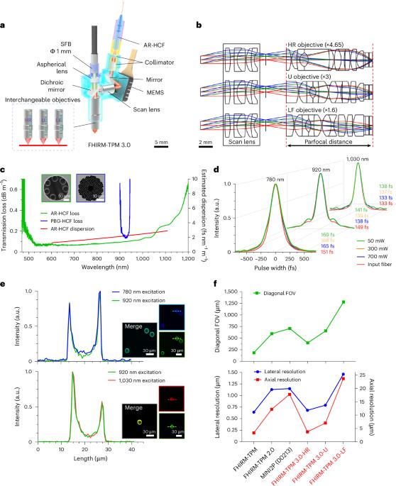
Here we present FHIRM-TPM 3.0, a 2.6 g miniature two-photon microscope capable of multicolor deep-brain imaging in freely behaving mice. The system was integrated with a broadband anti-resonant hollow-core fiber featuring low transmission loss, minimal dispersion from 700 nm to 1,060 nm and high tolerance of laser power. By correcting chromatic and spherical aberrations and optimizing the fluorescence collection aperture, we achieved cortical neuronal imaging at depths exceeding 820 μm and, using a GRIN lens, hippocampal Ca2+ imaging at single dendritic spine resolution. Moreover, we engineered three interchangeable parfocal objectives, allowing for a tenfold scalable field of view up to 1 × 0.8 mm², with lateral resolutions ranging from 0.68 μm to 1.46 μm. By multicolor imaging at excitation wavelengths of 780 nm, 920 nm and 1,030 nm, we investigated mitochondrial and cytosolic Ca2+ activities relative to the deposition of amyloid plaques in the cortex of awake APP/PS1 transgenic mice. Thus, FHIRM-TPM 3.0 provides a versatile imaging system suitable for diverse brain imaging scenarios. FHIRM-TPM 3.0 is a miniature microscope for multicolor two-photon imaging in freely moving mice. In addition to the multicolor imaging abilities achieved with the help of a specially designed optical fiber, the microscope is also compatible with multiple lenses for a choice of field of view and resolution.
期刊介绍:
Nature Methods is a monthly journal that focuses on publishing innovative methods and substantial enhancements to fundamental life sciences research techniques. Geared towards a diverse, interdisciplinary readership of researchers in academia and industry engaged in laboratory work, the journal offers new tools for research and emphasizes the immediate practical significance of the featured work. It publishes primary research papers and reviews recent technical and methodological advancements, with a particular interest in primary methods papers relevant to the biological and biomedical sciences. This includes methods rooted in chemistry with practical applications for studying biological problems.