{"title":"基于sbem的培养人细胞超微结构分析优化方案","authors":"Natalia Diak, Łukasz Chajec, Agnieszka Fus-Kujawa, Karolina Bajdak-Rusinek","doi":"10.3390/mps8040090","DOIUrl":null,"url":null,"abstract":"<p><p>Serial block-face scanning electron microscopy (SBEM) is a powerful technique for three-dimensional ultrastructural analysis of biological samples, though its application to in vitro cultured human cells remains underutilized. In this study, we present an optimized SBEM sample preparation protocol using human dermal fibroblasts and induced pluripotent stem cells (iPSCs). The method includes key modifications to the original protocol, such as using only glutaraldehyde for fixation and substituting the toxic cacodylate buffer with a less hazardous phosphate buffer. These adaptations result in excellent preservation of cellular ultrastructure, with high contrast and clarity, as validated by transmission electron microscopy (TEM). The loss of natural cell morphology resulted from fixation during passage, when cells formed a precipitate, rather than from fixation directly within the culture medium. The protocol is time-efficient, safe, and broadly applicable to both stem cells and differentiated cells cultured under 2D conditions, providing a valuable tool for ultrastructural analysis in diverse biomedical research settings.</p>","PeriodicalId":18715,"journal":{"name":"Methods and Protocols","volume":"8 4","pages":""},"PeriodicalIF":2.0000,"publicationDate":"2025-08-06","publicationTypes":"Journal Article","fieldsOfStudy":null,"isOpenAccess":false,"openAccessPdf":"https://www.ncbi.nlm.nih.gov/pmc/articles/PMC12388483/pdf/","citationCount":"0","resultStr":"{\"title\":\"An Optimized Protocol for SBEM-Based Ultrastructural Analysis of Cultured Human Cells.\",\"authors\":\"Natalia Diak, Łukasz Chajec, Agnieszka Fus-Kujawa, Karolina Bajdak-Rusinek\",\"doi\":\"10.3390/mps8040090\",\"DOIUrl\":null,\"url\":null,\"abstract\":\"<p><p>Serial block-face scanning electron microscopy (SBEM) is a powerful technique for three-dimensional ultrastructural analysis of biological samples, though its application to in vitro cultured human cells remains underutilized. In this study, we present an optimized SBEM sample preparation protocol using human dermal fibroblasts and induced pluripotent stem cells (iPSCs). The method includes key modifications to the original protocol, such as using only glutaraldehyde for fixation and substituting the toxic cacodylate buffer with a less hazardous phosphate buffer. These adaptations result in excellent preservation of cellular ultrastructure, with high contrast and clarity, as validated by transmission electron microscopy (TEM). The loss of natural cell morphology resulted from fixation during passage, when cells formed a precipitate, rather than from fixation directly within the culture medium. The protocol is time-efficient, safe, and broadly applicable to both stem cells and differentiated cells cultured under 2D conditions, providing a valuable tool for ultrastructural analysis in diverse biomedical research settings.</p>\",\"PeriodicalId\":18715,\"journal\":{\"name\":\"Methods and Protocols\",\"volume\":\"8 4\",\"pages\":\"\"},\"PeriodicalIF\":2.0000,\"publicationDate\":\"2025-08-06\",\"publicationTypes\":\"Journal Article\",\"fieldsOfStudy\":null,\"isOpenAccess\":false,\"openAccessPdf\":\"https://www.ncbi.nlm.nih.gov/pmc/articles/PMC12388483/pdf/\",\"citationCount\":\"0\",\"resultStr\":null,\"platform\":\"Semanticscholar\",\"paperid\":null,\"PeriodicalName\":\"Methods and Protocols\",\"FirstCategoryId\":\"1085\",\"ListUrlMain\":\"https://doi.org/10.3390/mps8040090\",\"RegionNum\":0,\"RegionCategory\":null,\"ArticlePicture\":[],\"TitleCN\":null,\"AbstractTextCN\":null,\"PMCID\":null,\"EPubDate\":\"\",\"PubModel\":\"\",\"JCR\":\"Q3\",\"JCRName\":\"BIOCHEMICAL RESEARCH METHODS\",\"Score\":null,\"Total\":0}","platform":"Semanticscholar","paperid":null,"PeriodicalName":"Methods and Protocols","FirstCategoryId":"1085","ListUrlMain":"https://doi.org/10.3390/mps8040090","RegionNum":0,"RegionCategory":null,"ArticlePicture":[],"TitleCN":null,"AbstractTextCN":null,"PMCID":null,"EPubDate":"","PubModel":"","JCR":"Q3","JCRName":"BIOCHEMICAL RESEARCH METHODS","Score":null,"Total":0}
An Optimized Protocol for SBEM-Based Ultrastructural Analysis of Cultured Human Cells.
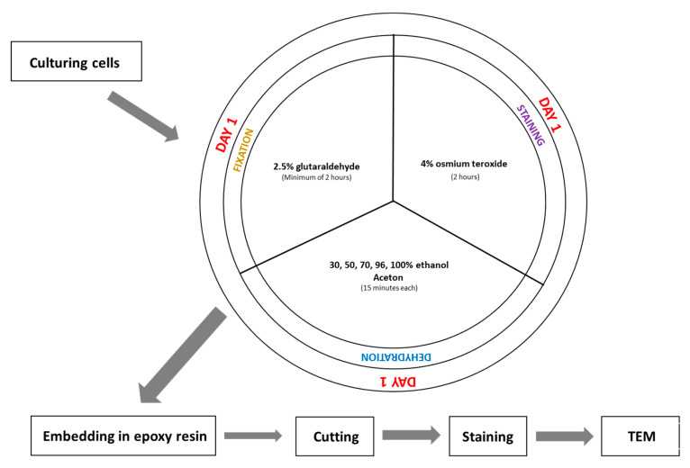
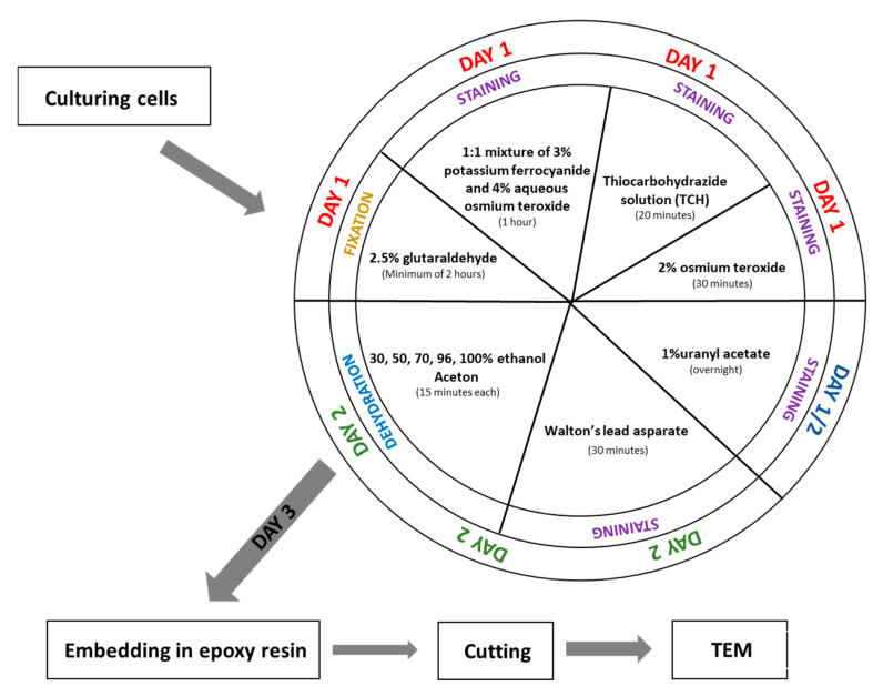
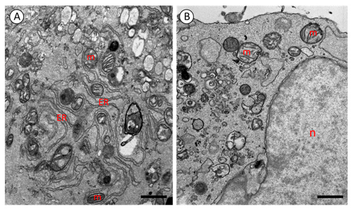
Serial block-face scanning electron microscopy (SBEM) is a powerful technique for three-dimensional ultrastructural analysis of biological samples, though its application to in vitro cultured human cells remains underutilized. In this study, we present an optimized SBEM sample preparation protocol using human dermal fibroblasts and induced pluripotent stem cells (iPSCs). The method includes key modifications to the original protocol, such as using only glutaraldehyde for fixation and substituting the toxic cacodylate buffer with a less hazardous phosphate buffer. These adaptations result in excellent preservation of cellular ultrastructure, with high contrast and clarity, as validated by transmission electron microscopy (TEM). The loss of natural cell morphology resulted from fixation during passage, when cells formed a precipitate, rather than from fixation directly within the culture medium. The protocol is time-efficient, safe, and broadly applicable to both stem cells and differentiated cells cultured under 2D conditions, providing a valuable tool for ultrastructural analysis in diverse biomedical research settings.

 求助内容:
求助内容: 应助结果提醒方式:


