{"title":"评价三维(3D)打印训练模型和定制腓骨模型在头颈部重建截骨和皮瓣植入训练中的教育效果。","authors":"Dharmik Gondalia, Mohit Parakh, Kunal Mokhale, Vineet Kumar, Ameya Bindu, Mayur Mantri, Saumya Mathews, Dushyant Jaiswal, Akshay Bhavke, Vinay Kant Shankhdhar","doi":"10.1186/s40902-025-00476-7","DOIUrl":null,"url":null,"abstract":"<p><strong>Background: </strong>Reconstructive surgery following head and neck cancer resection is inherently complex and technically demanding. Procedures such as osteotomy and flap inset involve a steep learning curve, yet opportunities for hands-on training are increasingly limited. Physical simulation using cost-effective, anatomically realistic models offers a promising solution. This study aimed to evaluate the educational value of 3D-printed training models and custom-made fibula models in enhancing surgical skills, supported by structured assessments and feedback.</p><p><strong>Methods: </strong>A hands-on workshop was conducted for 30 plastic surgery residents utilizing in-house 3D-printed models, created via fused deposition modeling (FDM), and acrylic-based fibula models. Participants performed simulated osteotomies and flap insets. Their performance was assessed using the 4-point Zwisch scale by two independent, blinded consultants. Additionally, a 6-item questionnaire was administered to capture self-reported improvements in anatomical understanding, surgical technique, and procedural planning. Pre- and post-training questionnaire scores were compared using Wilcoxon signed-rank test.</p><p><strong>Results: </strong>Post-training, the average questionnaire scores significantly improved from 12.03 ± 2.20 to 20.30 ± 1.56 (p < 0.01). The greatest improvement was noted in the participants' comprehension of surgical planning. Zwisch scale evaluations demonstrated a clear progression toward greater technical independence. Participants also expressed high satisfaction with the anatomical realism, durability, and affordability of the training models.</p><p><strong>Conclusion: </strong>Low-cost 3D-printed training models and custom-made fibula models represent an effective and reproducible training tool for developing technical skills in head and neck reconstructive surgery. Their ease of fabrication, affordability, and anatomical accuracy make them particularly valuable in resource-limited settings. These models offer significant educational utility and warrant integration into structured surgical training curriculum.</p>","PeriodicalId":18357,"journal":{"name":"Maxillofacial Plastic and Reconstructive Surgery","volume":"47 1","pages":"21"},"PeriodicalIF":2.8000,"publicationDate":"2025-08-20","publicationTypes":"Journal Article","fieldsOfStudy":null,"isOpenAccess":false,"openAccessPdf":"https://www.ncbi.nlm.nih.gov/pmc/articles/PMC12367569/pdf/","citationCount":"0","resultStr":"{\"title\":\"Evaluating educational effectiveness of three-dimensional (3D)-printed training models and custom-made fibula model for osteotomy and flap inset training in head and neck reconstruction.\",\"authors\":\"Dharmik Gondalia, Mohit Parakh, Kunal Mokhale, Vineet Kumar, Ameya Bindu, Mayur Mantri, Saumya Mathews, Dushyant Jaiswal, Akshay Bhavke, Vinay Kant Shankhdhar\",\"doi\":\"10.1186/s40902-025-00476-7\",\"DOIUrl\":null,\"url\":null,\"abstract\":\"<p><strong>Background: </strong>Reconstructive surgery following head and neck cancer resection is inherently complex and technically demanding. Procedures such as osteotomy and flap inset involve a steep learning curve, yet opportunities for hands-on training are increasingly limited. Physical simulation using cost-effective, anatomically realistic models offers a promising solution. This study aimed to evaluate the educational value of 3D-printed training models and custom-made fibula models in enhancing surgical skills, supported by structured assessments and feedback.</p><p><strong>Methods: </strong>A hands-on workshop was conducted for 30 plastic surgery residents utilizing in-house 3D-printed models, created via fused deposition modeling (FDM), and acrylic-based fibula models. Participants performed simulated osteotomies and flap insets. Their performance was assessed using the 4-point Zwisch scale by two independent, blinded consultants. Additionally, a 6-item questionnaire was administered to capture self-reported improvements in anatomical understanding, surgical technique, and procedural planning. Pre- and post-training questionnaire scores were compared using Wilcoxon signed-rank test.</p><p><strong>Results: </strong>Post-training, the average questionnaire scores significantly improved from 12.03 ± 2.20 to 20.30 ± 1.56 (p < 0.01). The greatest improvement was noted in the participants' comprehension of surgical planning. Zwisch scale evaluations demonstrated a clear progression toward greater technical independence. Participants also expressed high satisfaction with the anatomical realism, durability, and affordability of the training models.</p><p><strong>Conclusion: </strong>Low-cost 3D-printed training models and custom-made fibula models represent an effective and reproducible training tool for developing technical skills in head and neck reconstructive surgery. Their ease of fabrication, affordability, and anatomical accuracy make them particularly valuable in resource-limited settings. These models offer significant educational utility and warrant integration into structured surgical training curriculum.</p>\",\"PeriodicalId\":18357,\"journal\":{\"name\":\"Maxillofacial Plastic and Reconstructive Surgery\",\"volume\":\"47 1\",\"pages\":\"21\"},\"PeriodicalIF\":2.8000,\"publicationDate\":\"2025-08-20\",\"publicationTypes\":\"Journal Article\",\"fieldsOfStudy\":null,\"isOpenAccess\":false,\"openAccessPdf\":\"https://www.ncbi.nlm.nih.gov/pmc/articles/PMC12367569/pdf/\",\"citationCount\":\"0\",\"resultStr\":null,\"platform\":\"Semanticscholar\",\"paperid\":null,\"PeriodicalName\":\"Maxillofacial Plastic and Reconstructive Surgery\",\"FirstCategoryId\":\"1085\",\"ListUrlMain\":\"https://doi.org/10.1186/s40902-025-00476-7\",\"RegionNum\":0,\"RegionCategory\":null,\"ArticlePicture\":[],\"TitleCN\":null,\"AbstractTextCN\":null,\"PMCID\":null,\"EPubDate\":\"\",\"PubModel\":\"\",\"JCR\":\"Q2\",\"JCRName\":\"DENTISTRY, ORAL SURGERY & MEDICINE\",\"Score\":null,\"Total\":0}","platform":"Semanticscholar","paperid":null,"PeriodicalName":"Maxillofacial Plastic and Reconstructive Surgery","FirstCategoryId":"1085","ListUrlMain":"https://doi.org/10.1186/s40902-025-00476-7","RegionNum":0,"RegionCategory":null,"ArticlePicture":[],"TitleCN":null,"AbstractTextCN":null,"PMCID":null,"EPubDate":"","PubModel":"","JCR":"Q2","JCRName":"DENTISTRY, ORAL SURGERY & MEDICINE","Score":null,"Total":0}
Evaluating educational effectiveness of three-dimensional (3D)-printed training models and custom-made fibula model for osteotomy and flap inset training in head and neck reconstruction.
Background: Reconstructive surgery following head and neck cancer resection is inherently complex and technically demanding. Procedures such as osteotomy and flap inset involve a steep learning curve, yet opportunities for hands-on training are increasingly limited. Physical simulation using cost-effective, anatomically realistic models offers a promising solution. This study aimed to evaluate the educational value of 3D-printed training models and custom-made fibula models in enhancing surgical skills, supported by structured assessments and feedback.
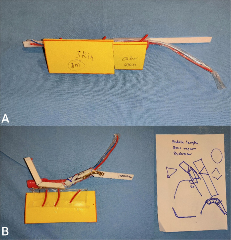
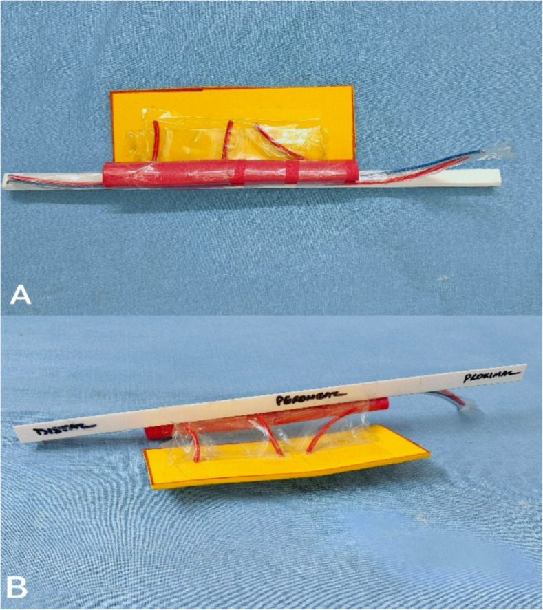
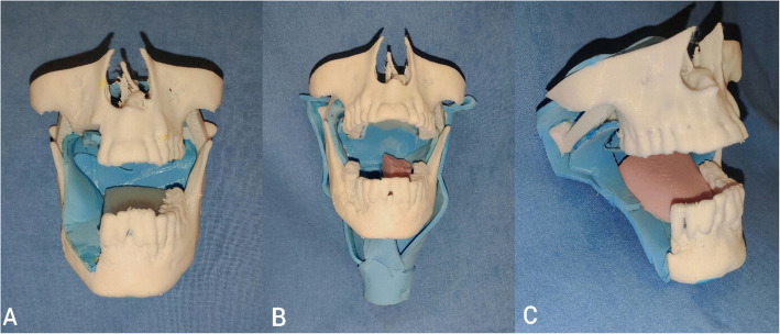
Methods: A hands-on workshop was conducted for 30 plastic surgery residents utilizing in-house 3D-printed models, created via fused deposition modeling (FDM), and acrylic-based fibula models. Participants performed simulated osteotomies and flap insets. Their performance was assessed using the 4-point Zwisch scale by two independent, blinded consultants. Additionally, a 6-item questionnaire was administered to capture self-reported improvements in anatomical understanding, surgical technique, and procedural planning. Pre- and post-training questionnaire scores were compared using Wilcoxon signed-rank test.
Results: Post-training, the average questionnaire scores significantly improved from 12.03 ± 2.20 to 20.30 ± 1.56 (p < 0.01). The greatest improvement was noted in the participants' comprehension of surgical planning. Zwisch scale evaluations demonstrated a clear progression toward greater technical independence. Participants also expressed high satisfaction with the anatomical realism, durability, and affordability of the training models.
Conclusion: Low-cost 3D-printed training models and custom-made fibula models represent an effective and reproducible training tool for developing technical skills in head and neck reconstructive surgery. Their ease of fabrication, affordability, and anatomical accuracy make them particularly valuable in resource-limited settings. These models offer significant educational utility and warrant integration into structured surgical training curriculum.

 求助内容:
求助内容: 应助结果提醒方式:


