Yeo Eun Han, Deuk Jae Sung, Hyun Yee Cho, Kyung Sook Yang, Jae Wook Park, Ki Choon Sim, Na Yeon Han, Beom Jin Park, Min Ju Kim
{"title":"膀胱浆细胞样尿路上皮癌的多参数MRI特征。","authors":"Yeo Eun Han, Deuk Jae Sung, Hyun Yee Cho, Kyung Sook Yang, Jae Wook Park, Ki Choon Sim, Na Yeon Han, Beom Jin Park, Min Ju Kim","doi":"10.3348/kjr.2025.0419","DOIUrl":null,"url":null,"abstract":"<p><strong>Objective: </strong>Plasmacytoid urothelial carcinoma (PUC) is a rare aggressive bladder cancer subtype with limited imaging data owing to its low incidence. This study aimed to report the characteristic features of PUC on multiparametric MRI (mpMRI).</p><p><strong>Materials and methods: </strong>We retrospectively analyzed 13 patients with histologically confirmed PUC who underwent preoperative mpMRI between January 2019 and August 2024. Two blinded radiologists independently assessed tumor size, morphology, signal intensity, apparent diffusion coefficient (ADC) values, dynamic contrast enhancement patterns, contrast enhancement features, and invasive characteristics. Vesical imaging-reporting and data system (VI-RADS) scores were recorded. Interobserver agreement was evaluated using the kappa statistic.</p><p><strong>Results: </strong>PUC predominantly exhibited diffuse (6/13, 46.2%) or localized (5/13, 38.5%) bladder wall thickening. Diffuse thickening was often associated with a linitis plastica-like appearance. On high b-value diffusion-weighted imaging (DWI), eight and seven cases depending on readers (61.5% and 53.8%, respectively) showed mild hyperintensity or isointensity, with a mean ADC value of 1.1 × 10⁻³ mm²/s. Dynamic contrast-enhanced MRI revealed progressive and prolonged enhancement in 10 cases (76.9%). VI-RADS scores ≥ 4 were observed in 11 cases (84.6%). Histopathological analysis showed that tumors with progressive and prolonged enhancement contained myxoid stroma and some fibrous tissue. Interobserver agreement was excellent for most imaging features, except for good agreement on DWI signal intensity.</p><p><strong>Conclusion: </strong>PUC demonstrates notable mpMRI features, including localized or diffuse wall thickening (often with a linitis plastica-like appearance), muscle-invasive and advanced disease, progressive and prolonged enhancement patterns, and mild hyperintensity or isointensity on high b-value DWI. These features, which are potentially linked to the myxoid stromal composition of the tumor, suggest that mpMRI may serve as a noninvasive diagnostic tool for this aggressive malignancy. However, further studies with larger cohorts are required to confirm these findings.</p>","PeriodicalId":17881,"journal":{"name":"Korean Journal of Radiology","volume":"26 9","pages":"832-840"},"PeriodicalIF":5.3000,"publicationDate":"2025-09-01","publicationTypes":"Journal Article","fieldsOfStudy":null,"isOpenAccess":false,"openAccessPdf":"https://www.ncbi.nlm.nih.gov/pmc/articles/PMC12394820/pdf/","citationCount":"0","resultStr":"{\"title\":\"Multiparametric MRI Features of Plasmacytoid Urothelial Carcinoma of the Urinary Bladder.\",\"authors\":\"Yeo Eun Han, Deuk Jae Sung, Hyun Yee Cho, Kyung Sook Yang, Jae Wook Park, Ki Choon Sim, Na Yeon Han, Beom Jin Park, Min Ju Kim\",\"doi\":\"10.3348/kjr.2025.0419\",\"DOIUrl\":null,\"url\":null,\"abstract\":\"<p><strong>Objective: </strong>Plasmacytoid urothelial carcinoma (PUC) is a rare aggressive bladder cancer subtype with limited imaging data owing to its low incidence. This study aimed to report the characteristic features of PUC on multiparametric MRI (mpMRI).</p><p><strong>Materials and methods: </strong>We retrospectively analyzed 13 patients with histologically confirmed PUC who underwent preoperative mpMRI between January 2019 and August 2024. Two blinded radiologists independently assessed tumor size, morphology, signal intensity, apparent diffusion coefficient (ADC) values, dynamic contrast enhancement patterns, contrast enhancement features, and invasive characteristics. Vesical imaging-reporting and data system (VI-RADS) scores were recorded. Interobserver agreement was evaluated using the kappa statistic.</p><p><strong>Results: </strong>PUC predominantly exhibited diffuse (6/13, 46.2%) or localized (5/13, 38.5%) bladder wall thickening. Diffuse thickening was often associated with a linitis plastica-like appearance. On high b-value diffusion-weighted imaging (DWI), eight and seven cases depending on readers (61.5% and 53.8%, respectively) showed mild hyperintensity or isointensity, with a mean ADC value of 1.1 × 10⁻³ mm²/s. Dynamic contrast-enhanced MRI revealed progressive and prolonged enhancement in 10 cases (76.9%). VI-RADS scores ≥ 4 were observed in 11 cases (84.6%). Histopathological analysis showed that tumors with progressive and prolonged enhancement contained myxoid stroma and some fibrous tissue. Interobserver agreement was excellent for most imaging features, except for good agreement on DWI signal intensity.</p><p><strong>Conclusion: </strong>PUC demonstrates notable mpMRI features, including localized or diffuse wall thickening (often with a linitis plastica-like appearance), muscle-invasive and advanced disease, progressive and prolonged enhancement patterns, and mild hyperintensity or isointensity on high b-value DWI. These features, which are potentially linked to the myxoid stromal composition of the tumor, suggest that mpMRI may serve as a noninvasive diagnostic tool for this aggressive malignancy. However, further studies with larger cohorts are required to confirm these findings.</p>\",\"PeriodicalId\":17881,\"journal\":{\"name\":\"Korean Journal of Radiology\",\"volume\":\"26 9\",\"pages\":\"832-840\"},\"PeriodicalIF\":5.3000,\"publicationDate\":\"2025-09-01\",\"publicationTypes\":\"Journal Article\",\"fieldsOfStudy\":null,\"isOpenAccess\":false,\"openAccessPdf\":\"https://www.ncbi.nlm.nih.gov/pmc/articles/PMC12394820/pdf/\",\"citationCount\":\"0\",\"resultStr\":null,\"platform\":\"Semanticscholar\",\"paperid\":null,\"PeriodicalName\":\"Korean Journal of Radiology\",\"FirstCategoryId\":\"3\",\"ListUrlMain\":\"https://doi.org/10.3348/kjr.2025.0419\",\"RegionNum\":2,\"RegionCategory\":\"医学\",\"ArticlePicture\":[],\"TitleCN\":null,\"AbstractTextCN\":null,\"PMCID\":null,\"EPubDate\":\"\",\"PubModel\":\"\",\"JCR\":\"Q1\",\"JCRName\":\"RADIOLOGY, NUCLEAR MEDICINE & MEDICAL IMAGING\",\"Score\":null,\"Total\":0}","platform":"Semanticscholar","paperid":null,"PeriodicalName":"Korean Journal of Radiology","FirstCategoryId":"3","ListUrlMain":"https://doi.org/10.3348/kjr.2025.0419","RegionNum":2,"RegionCategory":"医学","ArticlePicture":[],"TitleCN":null,"AbstractTextCN":null,"PMCID":null,"EPubDate":"","PubModel":"","JCR":"Q1","JCRName":"RADIOLOGY, NUCLEAR MEDICINE & MEDICAL IMAGING","Score":null,"Total":0}
Multiparametric MRI Features of Plasmacytoid Urothelial Carcinoma of the Urinary Bladder.
Objective: Plasmacytoid urothelial carcinoma (PUC) is a rare aggressive bladder cancer subtype with limited imaging data owing to its low incidence. This study aimed to report the characteristic features of PUC on multiparametric MRI (mpMRI).
Materials and methods: We retrospectively analyzed 13 patients with histologically confirmed PUC who underwent preoperative mpMRI between January 2019 and August 2024. Two blinded radiologists independently assessed tumor size, morphology, signal intensity, apparent diffusion coefficient (ADC) values, dynamic contrast enhancement patterns, contrast enhancement features, and invasive characteristics. Vesical imaging-reporting and data system (VI-RADS) scores were recorded. Interobserver agreement was evaluated using the kappa statistic.
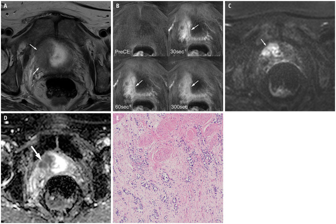
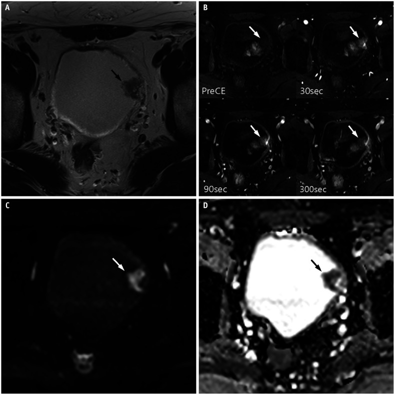
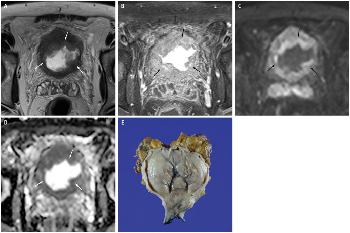
Results: PUC predominantly exhibited diffuse (6/13, 46.2%) or localized (5/13, 38.5%) bladder wall thickening. Diffuse thickening was often associated with a linitis plastica-like appearance. On high b-value diffusion-weighted imaging (DWI), eight and seven cases depending on readers (61.5% and 53.8%, respectively) showed mild hyperintensity or isointensity, with a mean ADC value of 1.1 × 10⁻³ mm²/s. Dynamic contrast-enhanced MRI revealed progressive and prolonged enhancement in 10 cases (76.9%). VI-RADS scores ≥ 4 were observed in 11 cases (84.6%). Histopathological analysis showed that tumors with progressive and prolonged enhancement contained myxoid stroma and some fibrous tissue. Interobserver agreement was excellent for most imaging features, except for good agreement on DWI signal intensity.
Conclusion: PUC demonstrates notable mpMRI features, including localized or diffuse wall thickening (often with a linitis plastica-like appearance), muscle-invasive and advanced disease, progressive and prolonged enhancement patterns, and mild hyperintensity or isointensity on high b-value DWI. These features, which are potentially linked to the myxoid stromal composition of the tumor, suggest that mpMRI may serve as a noninvasive diagnostic tool for this aggressive malignancy. However, further studies with larger cohorts are required to confirm these findings.
期刊介绍:
The inaugural issue of the Korean J Radiol came out in March 2000. Our journal aims to produce and propagate knowledge on radiologic imaging and related sciences.
A unique feature of the articles published in the Journal will be their reflection of global trends in radiology combined with an East-Asian perspective. Geographic differences in disease prevalence will be reflected in the contents of papers, and this will serve to enrich our body of knowledge.
World''s outstanding radiologists from many countries are serving as editorial board of our journal.

 求助内容:
求助内容: 应助结果提醒方式:


