Yue Ma, ChunGuo Wang, Han Yu, Benzhang Tao, Chao Gao, Gan Gao, Chao Xue
{"title":"腰椎退行性疾病骨盆长或短节段固定后坐位和站立位腰椎关节矢状位变化分析","authors":"Yue Ma, ChunGuo Wang, Han Yu, Benzhang Tao, Chao Gao, Gan Gao, Chao Xue","doi":"10.1186/s13018-025-06199-9","DOIUrl":null,"url":null,"abstract":"<p><strong>Background: </strong>The spine, pelvis, and joints maintain sagittal balance, which is often disrupted in lumbar degenerative diseases. While preoperative changes in sagittal alignment are well studied, postoperative adaptations, particularly following spinal fixation extending to the pelvis, are less understood. Therefore, the present study aimed to examine sagittal changes in the spine, pelvis, and joints in sitting and standing positions after short- or long-segment posterior spinal fixation extending to the pelvis.</p><p><strong>Methods: </strong>This cross-sectional study analyzed patients who underwent long- or short-segment instrumented fusion to the pelvis for lumbar degenerative disease at our hospital from June 2018 to October 2019. Patients were grouped based on the number of internal fixation segments, both short and long. Sagittal parameters were measured in standing and sitting positions and matched for sex, gender, height, weight, and other related parameters. Statistical analysis was performed using t-tests and Mann-Whitney U tests.</p><p><strong>Results: </strong>A total of 98 patients were included, of whom 55 were included in the long-segmengroup (31 men, 24 women; mean age of 63.1 ± 8.5 years). In the long-segment group, no significant changes were observed between standing and sitting positions (P > 0.05). In the short-segment group, significant changes were observed in the sacral vertical axis, pelvic tilt, sacral slope, thoracic kyphosis, lumbar lordosis, T1 pelvic angle, T1 spinopelvic inclination, acetabular tilt, and pelvic-femoral angle between the two positions (P < 0.05). The difference in pelvic femoral angle changes between the groups was also significant (P < 0.05).</p><p><strong>Conclusions: </strong>In the short-segment group, transitioning from standing to sitting leads to greater sagittal changes, including decreased lumbar lordosis and forward trunk lean, with smaller hip joints than in the long-segment internal fixation group.</p>","PeriodicalId":16629,"journal":{"name":"Journal of Orthopaedic Surgery and Research","volume":"20 1","pages":"803"},"PeriodicalIF":2.8000,"publicationDate":"2025-08-30","publicationTypes":"Journal Article","fieldsOfStudy":null,"isOpenAccess":false,"openAccessPdf":"https://www.ncbi.nlm.nih.gov/pmc/articles/PMC12398083/pdf/","citationCount":"0","resultStr":"{\"title\":\"Analysis of sagittal alignment changes in the spine-pelvis joint in sitting and standing positions after long- or short-segment fixation to the pelvis for lumbar degenerative diseases.\",\"authors\":\"Yue Ma, ChunGuo Wang, Han Yu, Benzhang Tao, Chao Gao, Gan Gao, Chao Xue\",\"doi\":\"10.1186/s13018-025-06199-9\",\"DOIUrl\":null,\"url\":null,\"abstract\":\"<p><strong>Background: </strong>The spine, pelvis, and joints maintain sagittal balance, which is often disrupted in lumbar degenerative diseases. While preoperative changes in sagittal alignment are well studied, postoperative adaptations, particularly following spinal fixation extending to the pelvis, are less understood. Therefore, the present study aimed to examine sagittal changes in the spine, pelvis, and joints in sitting and standing positions after short- or long-segment posterior spinal fixation extending to the pelvis.</p><p><strong>Methods: </strong>This cross-sectional study analyzed patients who underwent long- or short-segment instrumented fusion to the pelvis for lumbar degenerative disease at our hospital from June 2018 to October 2019. Patients were grouped based on the number of internal fixation segments, both short and long. Sagittal parameters were measured in standing and sitting positions and matched for sex, gender, height, weight, and other related parameters. Statistical analysis was performed using t-tests and Mann-Whitney U tests.</p><p><strong>Results: </strong>A total of 98 patients were included, of whom 55 were included in the long-segmengroup (31 men, 24 women; mean age of 63.1 ± 8.5 years). In the long-segment group, no significant changes were observed between standing and sitting positions (P > 0.05). In the short-segment group, significant changes were observed in the sacral vertical axis, pelvic tilt, sacral slope, thoracic kyphosis, lumbar lordosis, T1 pelvic angle, T1 spinopelvic inclination, acetabular tilt, and pelvic-femoral angle between the two positions (P < 0.05). The difference in pelvic femoral angle changes between the groups was also significant (P < 0.05).</p><p><strong>Conclusions: </strong>In the short-segment group, transitioning from standing to sitting leads to greater sagittal changes, including decreased lumbar lordosis and forward trunk lean, with smaller hip joints than in the long-segment internal fixation group.</p>\",\"PeriodicalId\":16629,\"journal\":{\"name\":\"Journal of Orthopaedic Surgery and Research\",\"volume\":\"20 1\",\"pages\":\"803\"},\"PeriodicalIF\":2.8000,\"publicationDate\":\"2025-08-30\",\"publicationTypes\":\"Journal Article\",\"fieldsOfStudy\":null,\"isOpenAccess\":false,\"openAccessPdf\":\"https://www.ncbi.nlm.nih.gov/pmc/articles/PMC12398083/pdf/\",\"citationCount\":\"0\",\"resultStr\":null,\"platform\":\"Semanticscholar\",\"paperid\":null,\"PeriodicalName\":\"Journal of Orthopaedic Surgery and Research\",\"FirstCategoryId\":\"3\",\"ListUrlMain\":\"https://doi.org/10.1186/s13018-025-06199-9\",\"RegionNum\":3,\"RegionCategory\":\"医学\",\"ArticlePicture\":[],\"TitleCN\":null,\"AbstractTextCN\":null,\"PMCID\":null,\"EPubDate\":\"\",\"PubModel\":\"\",\"JCR\":\"Q1\",\"JCRName\":\"ORTHOPEDICS\",\"Score\":null,\"Total\":0}","platform":"Semanticscholar","paperid":null,"PeriodicalName":"Journal of Orthopaedic Surgery and Research","FirstCategoryId":"3","ListUrlMain":"https://doi.org/10.1186/s13018-025-06199-9","RegionNum":3,"RegionCategory":"医学","ArticlePicture":[],"TitleCN":null,"AbstractTextCN":null,"PMCID":null,"EPubDate":"","PubModel":"","JCR":"Q1","JCRName":"ORTHOPEDICS","Score":null,"Total":0}
Analysis of sagittal alignment changes in the spine-pelvis joint in sitting and standing positions after long- or short-segment fixation to the pelvis for lumbar degenerative diseases.
Background: The spine, pelvis, and joints maintain sagittal balance, which is often disrupted in lumbar degenerative diseases. While preoperative changes in sagittal alignment are well studied, postoperative adaptations, particularly following spinal fixation extending to the pelvis, are less understood. Therefore, the present study aimed to examine sagittal changes in the spine, pelvis, and joints in sitting and standing positions after short- or long-segment posterior spinal fixation extending to the pelvis.
Methods: This cross-sectional study analyzed patients who underwent long- or short-segment instrumented fusion to the pelvis for lumbar degenerative disease at our hospital from June 2018 to October 2019. Patients were grouped based on the number of internal fixation segments, both short and long. Sagittal parameters were measured in standing and sitting positions and matched for sex, gender, height, weight, and other related parameters. Statistical analysis was performed using t-tests and Mann-Whitney U tests.
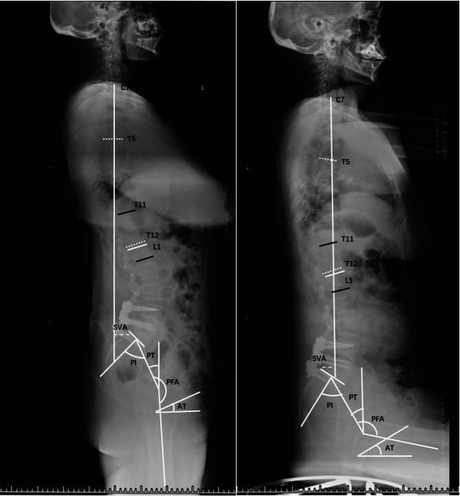
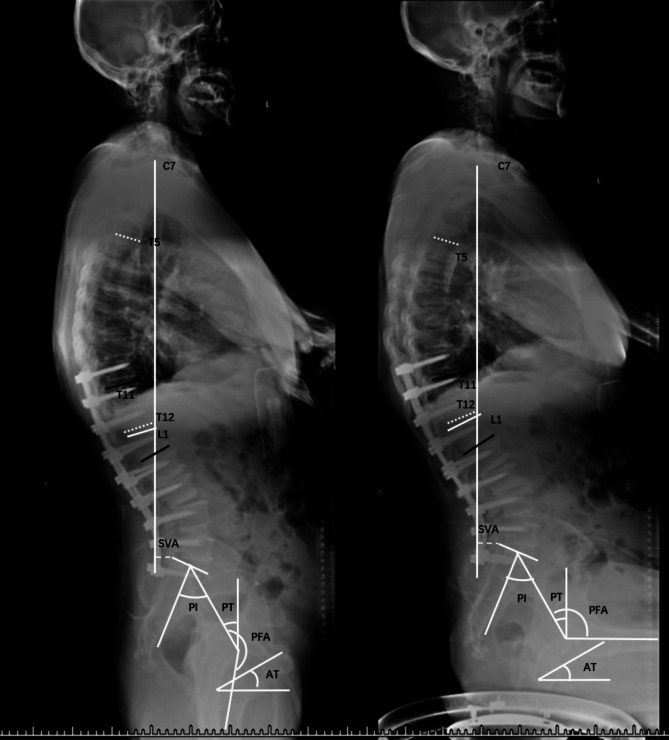
Results: A total of 98 patients were included, of whom 55 were included in the long-segmengroup (31 men, 24 women; mean age of 63.1 ± 8.5 years). In the long-segment group, no significant changes were observed between standing and sitting positions (P > 0.05). In the short-segment group, significant changes were observed in the sacral vertical axis, pelvic tilt, sacral slope, thoracic kyphosis, lumbar lordosis, T1 pelvic angle, T1 spinopelvic inclination, acetabular tilt, and pelvic-femoral angle between the two positions (P < 0.05). The difference in pelvic femoral angle changes between the groups was also significant (P < 0.05).
Conclusions: In the short-segment group, transitioning from standing to sitting leads to greater sagittal changes, including decreased lumbar lordosis and forward trunk lean, with smaller hip joints than in the long-segment internal fixation group.
期刊介绍:
Journal of Orthopaedic Surgery and Research is an open access journal that encompasses all aspects of clinical and basic research studies related to musculoskeletal issues.
Orthopaedic research is conducted at clinical and basic science levels. With the advancement of new technologies and the increasing expectation and demand from doctors and patients, we are witnessing an enormous growth in clinical orthopaedic research, particularly in the fields of traumatology, spinal surgery, joint replacement, sports medicine, musculoskeletal tumour management, hand microsurgery, foot and ankle surgery, paediatric orthopaedic, and orthopaedic rehabilitation. The involvement of basic science ranges from molecular, cellular, structural and functional perspectives to tissue engineering, gait analysis, automation and robotic surgery. Implant and biomaterial designs are new disciplines that complement clinical applications.
JOSR encourages the publication of multidisciplinary research with collaboration amongst clinicians and scientists from different disciplines, which will be the trend in the coming decades.

 求助内容:
求助内容: 应助结果提醒方式:


