Andrew M Hernandez, Anthony F Chen, Fatma Sen, Ana S Mitchell, Sarah E McKenney, Lorenzo Nardo, Craig K Abbey, Mohammad H Madani
{"title":"一项多读者、多病例研究,比较超分辨率和常规分辨率计算机断层扫描对肺结节特征的影响。","authors":"Andrew M Hernandez, Anthony F Chen, Fatma Sen, Ana S Mitchell, Sarah E McKenney, Lorenzo Nardo, Craig K Abbey, Mohammad H Madani","doi":"10.25259/JCIS_17_2025","DOIUrl":null,"url":null,"abstract":"<p><strong>Objectives: </strong>The objective of the study was to evaluate the efficacy of ultra-high-resolution computed tomography (UHRCT) in comparison to conventional resolution computed tomography (CT) for the characterization of lung nodules.</p><p><strong>Material and methods: </strong>104 non-contrast chest UHRCT scans (mean age of 66 years, 57 females) with pulmonary nodules were retrospectively collected (February-November 2022), and corresponding normal-resolution (NR) reconstructions were synthesized using a validated algorithm. Five blinded radiologists scored the following for each localized nodule in the ultra-high-resolution (UHR) and NR datasets: Margin clarity (5-point Likert scale), image quality \"IQ\" (3-point), density confidence (0-100%), and size (long/short axes). Image noise (voxel standard deviation) was calculated within the trachea. Differences between UHR and NR were tested using the Wilcoxon signed-rank test. Intrareader agreement was quantified with intraclass correlation coefficient (ICC), and ordinal association between margin clarity and IQ was quantified with Kendall's <i>τ</i> coefficient.</p><p><strong>Results: </strong>Margin clarity, IQ, and density confidence were significantly higher for UHR (<i>P</i> < 0.001). No significant differences between UHR and NR were observed in the variability (standard deviation across readers) for measuring long and short axes (<i>P</i> > 0.100). Intrareader agreement for UHR and NR was poor for margin clarity, IQ, and density confidences (ICC < 0.250) but moderate for short axes (ICC = 0.731) and good for long axes (ICC = 0.807). Ordinal association between margin clarity and IQ was moderate for UHR (<i>τ</i> = 0.566) and good for IQ (<i>τ</i> = 0.637). Image noise was significantly higher (<i>P</i> < 0.001) for UHR compared to NR.</p><p><strong>Conclusion: </strong>UHRCT offers significant improvements in the visualization of lung nodules compared to conventional resolution CT, albeit with an increase in image noise.</p>","PeriodicalId":15512,"journal":{"name":"Journal of Clinical Imaging Science","volume":"15 ","pages":"25"},"PeriodicalIF":1.3000,"publicationDate":"2025-07-08","publicationTypes":"Journal Article","fieldsOfStudy":null,"isOpenAccess":false,"openAccessPdf":"https://www.ncbi.nlm.nih.gov/pmc/articles/PMC12361665/pdf/","citationCount":"0","resultStr":"{\"title\":\"A multireader, multicase study comparing ultra-high-resolution and conventional-resolution computed tomography for lung nodule characterization.\",\"authors\":\"Andrew M Hernandez, Anthony F Chen, Fatma Sen, Ana S Mitchell, Sarah E McKenney, Lorenzo Nardo, Craig K Abbey, Mohammad H Madani\",\"doi\":\"10.25259/JCIS_17_2025\",\"DOIUrl\":null,\"url\":null,\"abstract\":\"<p><strong>Objectives: </strong>The objective of the study was to evaluate the efficacy of ultra-high-resolution computed tomography (UHRCT) in comparison to conventional resolution computed tomography (CT) for the characterization of lung nodules.</p><p><strong>Material and methods: </strong>104 non-contrast chest UHRCT scans (mean age of 66 years, 57 females) with pulmonary nodules were retrospectively collected (February-November 2022), and corresponding normal-resolution (NR) reconstructions were synthesized using a validated algorithm. Five blinded radiologists scored the following for each localized nodule in the ultra-high-resolution (UHR) and NR datasets: Margin clarity (5-point Likert scale), image quality \\\"IQ\\\" (3-point), density confidence (0-100%), and size (long/short axes). Image noise (voxel standard deviation) was calculated within the trachea. Differences between UHR and NR were tested using the Wilcoxon signed-rank test. Intrareader agreement was quantified with intraclass correlation coefficient (ICC), and ordinal association between margin clarity and IQ was quantified with Kendall's <i>τ</i> coefficient.</p><p><strong>Results: </strong>Margin clarity, IQ, and density confidence were significantly higher for UHR (<i>P</i> < 0.001). No significant differences between UHR and NR were observed in the variability (standard deviation across readers) for measuring long and short axes (<i>P</i> > 0.100). Intrareader agreement for UHR and NR was poor for margin clarity, IQ, and density confidences (ICC < 0.250) but moderate for short axes (ICC = 0.731) and good for long axes (ICC = 0.807). Ordinal association between margin clarity and IQ was moderate for UHR (<i>τ</i> = 0.566) and good for IQ (<i>τ</i> = 0.637). Image noise was significantly higher (<i>P</i> < 0.001) for UHR compared to NR.</p><p><strong>Conclusion: </strong>UHRCT offers significant improvements in the visualization of lung nodules compared to conventional resolution CT, albeit with an increase in image noise.</p>\",\"PeriodicalId\":15512,\"journal\":{\"name\":\"Journal of Clinical Imaging Science\",\"volume\":\"15 \",\"pages\":\"25\"},\"PeriodicalIF\":1.3000,\"publicationDate\":\"2025-07-08\",\"publicationTypes\":\"Journal Article\",\"fieldsOfStudy\":null,\"isOpenAccess\":false,\"openAccessPdf\":\"https://www.ncbi.nlm.nih.gov/pmc/articles/PMC12361665/pdf/\",\"citationCount\":\"0\",\"resultStr\":null,\"platform\":\"Semanticscholar\",\"paperid\":null,\"PeriodicalName\":\"Journal of Clinical Imaging Science\",\"FirstCategoryId\":\"1085\",\"ListUrlMain\":\"https://doi.org/10.25259/JCIS_17_2025\",\"RegionNum\":0,\"RegionCategory\":null,\"ArticlePicture\":[],\"TitleCN\":null,\"AbstractTextCN\":null,\"PMCID\":null,\"EPubDate\":\"2025/1/1 0:00:00\",\"PubModel\":\"eCollection\",\"JCR\":\"Q3\",\"JCRName\":\"RADIOLOGY, NUCLEAR MEDICINE & MEDICAL IMAGING\",\"Score\":null,\"Total\":0}","platform":"Semanticscholar","paperid":null,"PeriodicalName":"Journal of Clinical Imaging Science","FirstCategoryId":"1085","ListUrlMain":"https://doi.org/10.25259/JCIS_17_2025","RegionNum":0,"RegionCategory":null,"ArticlePicture":[],"TitleCN":null,"AbstractTextCN":null,"PMCID":null,"EPubDate":"2025/1/1 0:00:00","PubModel":"eCollection","JCR":"Q3","JCRName":"RADIOLOGY, NUCLEAR MEDICINE & MEDICAL IMAGING","Score":null,"Total":0}
A multireader, multicase study comparing ultra-high-resolution and conventional-resolution computed tomography for lung nodule characterization.
Objectives: The objective of the study was to evaluate the efficacy of ultra-high-resolution computed tomography (UHRCT) in comparison to conventional resolution computed tomography (CT) for the characterization of lung nodules.
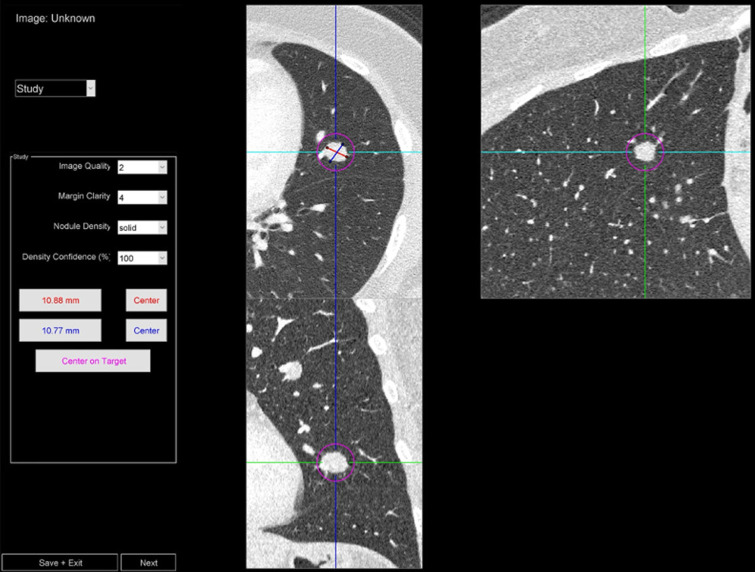
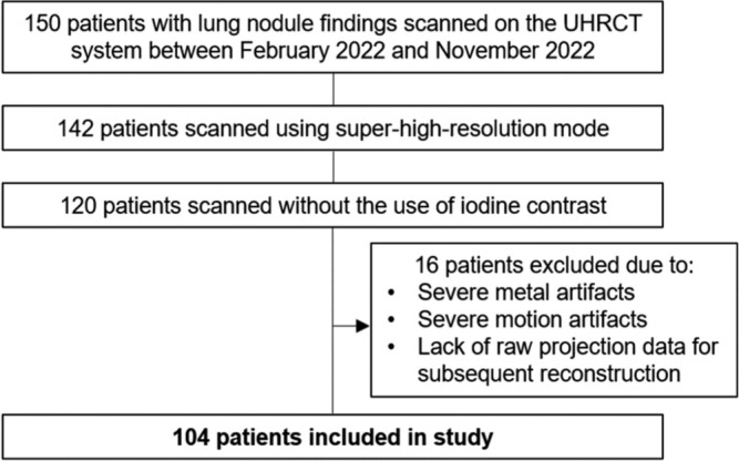
Material and methods: 104 non-contrast chest UHRCT scans (mean age of 66 years, 57 females) with pulmonary nodules were retrospectively collected (February-November 2022), and corresponding normal-resolution (NR) reconstructions were synthesized using a validated algorithm. Five blinded radiologists scored the following for each localized nodule in the ultra-high-resolution (UHR) and NR datasets: Margin clarity (5-point Likert scale), image quality "IQ" (3-point), density confidence (0-100%), and size (long/short axes). Image noise (voxel standard deviation) was calculated within the trachea. Differences between UHR and NR were tested using the Wilcoxon signed-rank test. Intrareader agreement was quantified with intraclass correlation coefficient (ICC), and ordinal association between margin clarity and IQ was quantified with Kendall's τ coefficient.
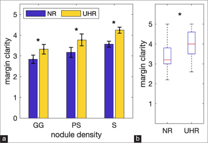
Results: Margin clarity, IQ, and density confidence were significantly higher for UHR (P < 0.001). No significant differences between UHR and NR were observed in the variability (standard deviation across readers) for measuring long and short axes (P > 0.100). Intrareader agreement for UHR and NR was poor for margin clarity, IQ, and density confidences (ICC < 0.250) but moderate for short axes (ICC = 0.731) and good for long axes (ICC = 0.807). Ordinal association between margin clarity and IQ was moderate for UHR (τ = 0.566) and good for IQ (τ = 0.637). Image noise was significantly higher (P < 0.001) for UHR compared to NR.
Conclusion: UHRCT offers significant improvements in the visualization of lung nodules compared to conventional resolution CT, albeit with an increase in image noise.
期刊介绍:
The Journal of Clinical Imaging Science (JCIS) is an open access peer-reviewed journal committed to publishing high-quality articles in the field of Imaging Science. The journal aims to present Imaging Science and relevant clinical information in an understandable and useful format. The journal is owned and published by the Scientific Scholar. Audience Our audience includes Radiologists, Researchers, Clinicians, medical professionals and students. Review process JCIS has a highly rigorous peer-review process that makes sure that manuscripts are scientifically accurate, relevant, novel and important. Authors disclose all conflicts, affiliations and financial associations such that the published content is not biased.

 求助内容:
求助内容: 应助结果提醒方式:


