{"title":"磁共振成像在急性心肌梗死合并心内出血患者治疗中的应用。","authors":"Xiao-Long Mi, Li-Li Zhang, Yan-Hui Zhang, Zheng Xu, Peng-Fei Ding, Dong Sun","doi":"10.1186/s13019-025-03574-9","DOIUrl":null,"url":null,"abstract":"<p><strong>Background: </strong>We assessed the diagnostic efficacy of magnetic resonance imaging (MRI) in patients with acute myocardial infarction (AMI).</p><p><strong>Methods: </strong>In this study, 116 patients with acute myocardial infarction (AMI) underwent direct PCI intervention, admitted to our hospital between January 2018 and January 2021 were selected. Based on the presence of intramyocardial hemorrhage (IMH), they were divided into the IMH group and the non-IMH group. There were 46 cases in the IMH group and 70 cases in the non-IMH group. All patients underwent cardiac magnetic resonance imaging (CMR) for detection. CMR was used to detect IMH and non-IMH infarction sites. Cardiac indicators of IMH and non-IMH were compared using CMR and echocardiography (ECHO). The diagnostic efficacy of MRI in patients with AMI who had myocardial hemorrhage was compared by generating receiver operating characteristic (ROC) curves.</p><p><strong>Results: </strong>The incidence of infarction sites was significantly higher in the IMH group than in the non-IMH group (all P < 0.05); myocardial detection results revealed a significantly higher incidence of ventricular aneurysm and pericardial fluid inclusion in the IMH group than in the non-IMH group (all P < 0.05); CMR evaluation revealed that the infarction size/left ventricular (IS/LV) volume percentage, patients with microvascular obstruction (MVO), and MVO/LV volume percentage were significantly higher in the IMH group than in the non-IMH group (all P < 0.05); global circumferential strain (GCS), global radial strain (GRS), and global longitudinal strain (GLS) in the IMH group were significantly lower than those in the non-IMH group (all P < 0.05); was both groups underwent echocardiography after percutaneous coronary intervention (PCI). The results indicated a significant decrease in left ventricular ejection fraction (LVEF) and a significant increase in left ventricular end-diastolic dimension (LVEDd) and IS/LV volume percentage in the IMH group compared to the non-IMH group (all P < 0.05); the area under the ROC curve of MRI for patients with AMI who had intramyocardial hemorrhage was 0.869, with high specificity and sensitivity; the sensitivity was 87.00, and the specificity was 85.00.</p><p><strong>Conclusion: </strong>MRI can detect myocardial hemorrhage in patients with AMI after PCI, which suggests significant clinical diagnostic value and is worthy of utilization in clinical practice.</p>","PeriodicalId":15201,"journal":{"name":"Journal of Cardiothoracic Surgery","volume":"20 1","pages":"348"},"PeriodicalIF":1.5000,"publicationDate":"2025-08-29","publicationTypes":"Journal Article","fieldsOfStudy":null,"isOpenAccess":false,"openAccessPdf":"https://www.ncbi.nlm.nih.gov/pmc/articles/PMC12395912/pdf/","citationCount":"0","resultStr":"{\"title\":\"Utilization of magnetic resonance imaging in the treatment of patients with acute myocardial infarction and intramyocardial hemorrhage.\",\"authors\":\"Xiao-Long Mi, Li-Li Zhang, Yan-Hui Zhang, Zheng Xu, Peng-Fei Ding, Dong Sun\",\"doi\":\"10.1186/s13019-025-03574-9\",\"DOIUrl\":null,\"url\":null,\"abstract\":\"<p><strong>Background: </strong>We assessed the diagnostic efficacy of magnetic resonance imaging (MRI) in patients with acute myocardial infarction (AMI).</p><p><strong>Methods: </strong>In this study, 116 patients with acute myocardial infarction (AMI) underwent direct PCI intervention, admitted to our hospital between January 2018 and January 2021 were selected. Based on the presence of intramyocardial hemorrhage (IMH), they were divided into the IMH group and the non-IMH group. There were 46 cases in the IMH group and 70 cases in the non-IMH group. All patients underwent cardiac magnetic resonance imaging (CMR) for detection. CMR was used to detect IMH and non-IMH infarction sites. Cardiac indicators of IMH and non-IMH were compared using CMR and echocardiography (ECHO). The diagnostic efficacy of MRI in patients with AMI who had myocardial hemorrhage was compared by generating receiver operating characteristic (ROC) curves.</p><p><strong>Results: </strong>The incidence of infarction sites was significantly higher in the IMH group than in the non-IMH group (all P < 0.05); myocardial detection results revealed a significantly higher incidence of ventricular aneurysm and pericardial fluid inclusion in the IMH group than in the non-IMH group (all P < 0.05); CMR evaluation revealed that the infarction size/left ventricular (IS/LV) volume percentage, patients with microvascular obstruction (MVO), and MVO/LV volume percentage were significantly higher in the IMH group than in the non-IMH group (all P < 0.05); global circumferential strain (GCS), global radial strain (GRS), and global longitudinal strain (GLS) in the IMH group were significantly lower than those in the non-IMH group (all P < 0.05); was both groups underwent echocardiography after percutaneous coronary intervention (PCI). The results indicated a significant decrease in left ventricular ejection fraction (LVEF) and a significant increase in left ventricular end-diastolic dimension (LVEDd) and IS/LV volume percentage in the IMH group compared to the non-IMH group (all P < 0.05); the area under the ROC curve of MRI for patients with AMI who had intramyocardial hemorrhage was 0.869, with high specificity and sensitivity; the sensitivity was 87.00, and the specificity was 85.00.</p><p><strong>Conclusion: </strong>MRI can detect myocardial hemorrhage in patients with AMI after PCI, which suggests significant clinical diagnostic value and is worthy of utilization in clinical practice.</p>\",\"PeriodicalId\":15201,\"journal\":{\"name\":\"Journal of Cardiothoracic Surgery\",\"volume\":\"20 1\",\"pages\":\"348\"},\"PeriodicalIF\":1.5000,\"publicationDate\":\"2025-08-29\",\"publicationTypes\":\"Journal Article\",\"fieldsOfStudy\":null,\"isOpenAccess\":false,\"openAccessPdf\":\"https://www.ncbi.nlm.nih.gov/pmc/articles/PMC12395912/pdf/\",\"citationCount\":\"0\",\"resultStr\":null,\"platform\":\"Semanticscholar\",\"paperid\":null,\"PeriodicalName\":\"Journal of Cardiothoracic Surgery\",\"FirstCategoryId\":\"3\",\"ListUrlMain\":\"https://doi.org/10.1186/s13019-025-03574-9\",\"RegionNum\":4,\"RegionCategory\":\"医学\",\"ArticlePicture\":[],\"TitleCN\":null,\"AbstractTextCN\":null,\"PMCID\":null,\"EPubDate\":\"\",\"PubModel\":\"\",\"JCR\":\"Q3\",\"JCRName\":\"CARDIAC & CARDIOVASCULAR SYSTEMS\",\"Score\":null,\"Total\":0}","platform":"Semanticscholar","paperid":null,"PeriodicalName":"Journal of Cardiothoracic Surgery","FirstCategoryId":"3","ListUrlMain":"https://doi.org/10.1186/s13019-025-03574-9","RegionNum":4,"RegionCategory":"医学","ArticlePicture":[],"TitleCN":null,"AbstractTextCN":null,"PMCID":null,"EPubDate":"","PubModel":"","JCR":"Q3","JCRName":"CARDIAC & CARDIOVASCULAR SYSTEMS","Score":null,"Total":0}
Utilization of magnetic resonance imaging in the treatment of patients with acute myocardial infarction and intramyocardial hemorrhage.
Background: We assessed the diagnostic efficacy of magnetic resonance imaging (MRI) in patients with acute myocardial infarction (AMI).
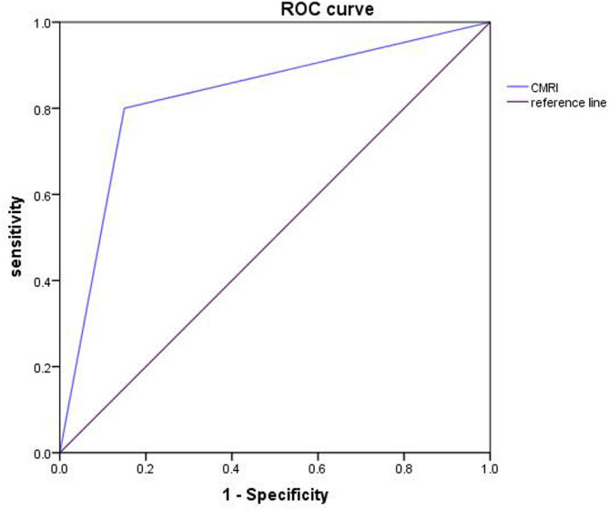
Methods: In this study, 116 patients with acute myocardial infarction (AMI) underwent direct PCI intervention, admitted to our hospital between January 2018 and January 2021 were selected. Based on the presence of intramyocardial hemorrhage (IMH), they were divided into the IMH group and the non-IMH group. There were 46 cases in the IMH group and 70 cases in the non-IMH group. All patients underwent cardiac magnetic resonance imaging (CMR) for detection. CMR was used to detect IMH and non-IMH infarction sites. Cardiac indicators of IMH and non-IMH were compared using CMR and echocardiography (ECHO). The diagnostic efficacy of MRI in patients with AMI who had myocardial hemorrhage was compared by generating receiver operating characteristic (ROC) curves.
Results: The incidence of infarction sites was significantly higher in the IMH group than in the non-IMH group (all P < 0.05); myocardial detection results revealed a significantly higher incidence of ventricular aneurysm and pericardial fluid inclusion in the IMH group than in the non-IMH group (all P < 0.05); CMR evaluation revealed that the infarction size/left ventricular (IS/LV) volume percentage, patients with microvascular obstruction (MVO), and MVO/LV volume percentage were significantly higher in the IMH group than in the non-IMH group (all P < 0.05); global circumferential strain (GCS), global radial strain (GRS), and global longitudinal strain (GLS) in the IMH group were significantly lower than those in the non-IMH group (all P < 0.05); was both groups underwent echocardiography after percutaneous coronary intervention (PCI). The results indicated a significant decrease in left ventricular ejection fraction (LVEF) and a significant increase in left ventricular end-diastolic dimension (LVEDd) and IS/LV volume percentage in the IMH group compared to the non-IMH group (all P < 0.05); the area under the ROC curve of MRI for patients with AMI who had intramyocardial hemorrhage was 0.869, with high specificity and sensitivity; the sensitivity was 87.00, and the specificity was 85.00.
Conclusion: MRI can detect myocardial hemorrhage in patients with AMI after PCI, which suggests significant clinical diagnostic value and is worthy of utilization in clinical practice.
期刊介绍:
Journal of Cardiothoracic Surgery is an open access journal that encompasses all aspects of research in the field of Cardiology, and Cardiothoracic and Vascular Surgery. The journal publishes original scientific research documenting clinical and experimental advances in cardiac, vascular and thoracic surgery, and related fields.
Topics of interest include surgical techniques, survival rates, surgical complications and their outcomes; along with basic sciences, pediatric conditions, transplantations and clinical trials.
Journal of Cardiothoracic Surgery is of interest to cardiothoracic and vascular surgeons, cardiothoracic anaesthesiologists, cardiologists, chest physicians, and allied health professionals.
 求助内容:
求助内容: 应助结果提醒方式:


