Kel Vin Woo, Philip T Levy, Carla J Weinheimer, Amanda L Hauck, Aaron Hamvas, David M Ornitz, Attila Kovacs, Gautam K Singh
{"title":"新生儿缺氧小鼠肺血流动力学和右心室功能的超声心动图评价。","authors":"Kel Vin Woo, Philip T Levy, Carla J Weinheimer, Amanda L Hauck, Aaron Hamvas, David M Ornitz, Attila Kovacs, Gautam K Singh","doi":"10.3390/jcdd12080316","DOIUrl":null,"url":null,"abstract":"<p><p><b>Background:</b> Right heart catheterization (RHC) is the gold-standard for diagnosis of pulmonary hypertension (PH) but is a terminal procedure in neonatal mice. The objective was to validate echocardiographic measures of PH to establish the diagnostic capability against pulmonary vascular histology in neonatal mice. <b>Methods:</b> Adult mice, exposed to hypoxia or normoxia, were assessed by echocardiography and RHC to evaluate right ventricle (RV) morphometry and function. Echocardiographic measures identified in adult mice were then used to evaluate PH characteristics in hypoxia-exposed neonatal mice. Physiological parameters were compared to histopathology in all mice. <b>Results:</b> Hypoxia-challenged adult mice developed PH with RHC, demonstrating confirmed elevated RV systolic pressure (RVSP), RV hypertrophy, and increased cross-sectional area and neomuscularization of pulmonary vessels. Echocardiography-derived RV free wall (RVFW) thickness correlated with RV mass. Tricuspid valve annulus tissue Doppler imaging (TV TDI), tricuspid annular plane systolic excursion (TAPSE), pulmonary artery acceleration measures (PAAT), and TAPSE × PAAT (a measure of RV work) all correlated with RVSP determined by RHC. In neonatal mice exposed to hypoxia, PAAT, TV TDI, TAPSE, and TAPSE × PAAT were decreased and RVFW thickness was increased, correlating with the histologic phenotype of PH. <b>Conclusions:</b> Echocardiographic indices of RV morphology and function provide reliable estimates of invasive RV hemodynamics in hypoxia-induced PH.</p>","PeriodicalId":15197,"journal":{"name":"Journal of Cardiovascular Development and Disease","volume":"12 8","pages":""},"PeriodicalIF":2.3000,"publicationDate":"2025-08-19","publicationTypes":"Journal Article","fieldsOfStudy":null,"isOpenAccess":false,"openAccessPdf":"https://www.ncbi.nlm.nih.gov/pmc/articles/PMC12386579/pdf/","citationCount":"0","resultStr":"{\"title\":\"Echocardiographic Assessment of Pulmonary Hemodynamics and Right Ventricular Performance in Neonatal Murine Hypoxia.\",\"authors\":\"Kel Vin Woo, Philip T Levy, Carla J Weinheimer, Amanda L Hauck, Aaron Hamvas, David M Ornitz, Attila Kovacs, Gautam K Singh\",\"doi\":\"10.3390/jcdd12080316\",\"DOIUrl\":null,\"url\":null,\"abstract\":\"<p><p><b>Background:</b> Right heart catheterization (RHC) is the gold-standard for diagnosis of pulmonary hypertension (PH) but is a terminal procedure in neonatal mice. The objective was to validate echocardiographic measures of PH to establish the diagnostic capability against pulmonary vascular histology in neonatal mice. <b>Methods:</b> Adult mice, exposed to hypoxia or normoxia, were assessed by echocardiography and RHC to evaluate right ventricle (RV) morphometry and function. Echocardiographic measures identified in adult mice were then used to evaluate PH characteristics in hypoxia-exposed neonatal mice. Physiological parameters were compared to histopathology in all mice. <b>Results:</b> Hypoxia-challenged adult mice developed PH with RHC, demonstrating confirmed elevated RV systolic pressure (RVSP), RV hypertrophy, and increased cross-sectional area and neomuscularization of pulmonary vessels. Echocardiography-derived RV free wall (RVFW) thickness correlated with RV mass. Tricuspid valve annulus tissue Doppler imaging (TV TDI), tricuspid annular plane systolic excursion (TAPSE), pulmonary artery acceleration measures (PAAT), and TAPSE × PAAT (a measure of RV work) all correlated with RVSP determined by RHC. In neonatal mice exposed to hypoxia, PAAT, TV TDI, TAPSE, and TAPSE × PAAT were decreased and RVFW thickness was increased, correlating with the histologic phenotype of PH. <b>Conclusions:</b> Echocardiographic indices of RV morphology and function provide reliable estimates of invasive RV hemodynamics in hypoxia-induced PH.</p>\",\"PeriodicalId\":15197,\"journal\":{\"name\":\"Journal of Cardiovascular Development and Disease\",\"volume\":\"12 8\",\"pages\":\"\"},\"PeriodicalIF\":2.3000,\"publicationDate\":\"2025-08-19\",\"publicationTypes\":\"Journal Article\",\"fieldsOfStudy\":null,\"isOpenAccess\":false,\"openAccessPdf\":\"https://www.ncbi.nlm.nih.gov/pmc/articles/PMC12386579/pdf/\",\"citationCount\":\"0\",\"resultStr\":null,\"platform\":\"Semanticscholar\",\"paperid\":null,\"PeriodicalName\":\"Journal of Cardiovascular Development and Disease\",\"FirstCategoryId\":\"3\",\"ListUrlMain\":\"https://doi.org/10.3390/jcdd12080316\",\"RegionNum\":4,\"RegionCategory\":\"医学\",\"ArticlePicture\":[],\"TitleCN\":null,\"AbstractTextCN\":null,\"PMCID\":null,\"EPubDate\":\"\",\"PubModel\":\"\",\"JCR\":\"Q2\",\"JCRName\":\"CARDIAC & CARDIOVASCULAR SYSTEMS\",\"Score\":null,\"Total\":0}","platform":"Semanticscholar","paperid":null,"PeriodicalName":"Journal of Cardiovascular Development and Disease","FirstCategoryId":"3","ListUrlMain":"https://doi.org/10.3390/jcdd12080316","RegionNum":4,"RegionCategory":"医学","ArticlePicture":[],"TitleCN":null,"AbstractTextCN":null,"PMCID":null,"EPubDate":"","PubModel":"","JCR":"Q2","JCRName":"CARDIAC & CARDIOVASCULAR SYSTEMS","Score":null,"Total":0}
Echocardiographic Assessment of Pulmonary Hemodynamics and Right Ventricular Performance in Neonatal Murine Hypoxia.
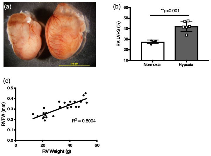
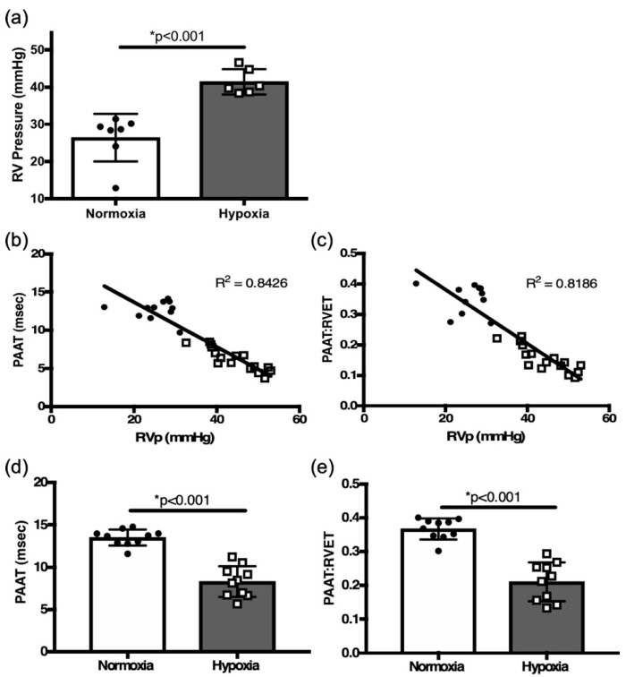
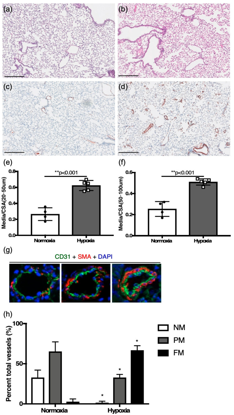
Background: Right heart catheterization (RHC) is the gold-standard for diagnosis of pulmonary hypertension (PH) but is a terminal procedure in neonatal mice. The objective was to validate echocardiographic measures of PH to establish the diagnostic capability against pulmonary vascular histology in neonatal mice. Methods: Adult mice, exposed to hypoxia or normoxia, were assessed by echocardiography and RHC to evaluate right ventricle (RV) morphometry and function. Echocardiographic measures identified in adult mice were then used to evaluate PH characteristics in hypoxia-exposed neonatal mice. Physiological parameters were compared to histopathology in all mice. Results: Hypoxia-challenged adult mice developed PH with RHC, demonstrating confirmed elevated RV systolic pressure (RVSP), RV hypertrophy, and increased cross-sectional area and neomuscularization of pulmonary vessels. Echocardiography-derived RV free wall (RVFW) thickness correlated with RV mass. Tricuspid valve annulus tissue Doppler imaging (TV TDI), tricuspid annular plane systolic excursion (TAPSE), pulmonary artery acceleration measures (PAAT), and TAPSE × PAAT (a measure of RV work) all correlated with RVSP determined by RHC. In neonatal mice exposed to hypoxia, PAAT, TV TDI, TAPSE, and TAPSE × PAAT were decreased and RVFW thickness was increased, correlating with the histologic phenotype of PH. Conclusions: Echocardiographic indices of RV morphology and function provide reliable estimates of invasive RV hemodynamics in hypoxia-induced PH.

 求助内容:
求助内容: 应助结果提醒方式:


