Minh Dien Tran, Sheetal Maria Rajan, Hien Chi Ngo, Amr Fawzy
{"title":"用于生物膜清创的高强度聚焦超声,使用钛附着的变形链球菌的体外概念验证。","authors":"Minh Dien Tran, Sheetal Maria Rajan, Hien Chi Ngo, Amr Fawzy","doi":"10.1186/s40729-025-00645-3","DOIUrl":null,"url":null,"abstract":"<p><strong>Introduction: </strong>Peri-implantitis (PI) is a biofilm-related condition driven by bacterial colonization on dental implant surfaces, leading to inflammation of the peri-implant connective tissue and progressive bone loss. Despite advancements, effective strategies for eradicating these biofilms remain elusive. While high-intensity focused ultrasound (HIFU) has been popularized in medicine, its effects on dental implant-attached biofilms remain unclear. This study presents in vitro findings on the effects of HIFU treatment on titanium (Ti)-attached Streptococcus mutans (S. mutans) biofilms and evaluates its impacts on the surface roughness and chemical composition of the Ti disc substrates.</p><p><strong>Methods: </strong>To optimise the HIFU parameters, four quadrants of a pair of Ti discs [machined (M) and alumina grit blasted (AB)] were marked using laser etching (MD Waterlase, US). HIFU beams, generated by a 254 kHz transducer and operated at intensities of 0 W, 10 W, 20 W, and 30 W, were applied to each quadrant for 2 min (min) in a water medium. The roughness of the treated surfaces was measured using Atomic Force Microscopy (AFM), and the surface composition was analyzed using Scanning Electron Microscope-Energy Dispersive Spectrometry (SEM-EDS). To investigate the biofilm debridement, 10-day-old S. mutans cultures were grown on 20 pairs of similar Ti discs, and then the optimized HIFU intensity of 20W was applied to five test pairs. Qualitative analyses were performed using a Dual Fluorescence/Reflection Confocal Laser Scanning Microscope (FRCLSM) and SEM imaging. Quantitative data on cell viability were collected using crystal violet (CV), (3-[4,5-dimethylthiazol-2-yl]-2,5 2,5-diphenyl tetrazolium bromide) (MTT), and flow cytometry (FCM) assays. Data from these test conditions were analyzed alongside cultures on biofilms that were untreated (control). Statistical data were calculated using ANOVA and appropriate t-tests for repeated measures.</p><p><strong>Results: </strong>The surface roughness of AB Ti discs showed a highest and significant increase (p < 0.05) following HIFU exposure at 20 W through three roughness parameters (Sa, Sq, and Sdr), compared to the controls (1207 nm, 1455 nm, 62% compared to 842 nm, 1042 nm and 30% respectively). This optimized HIFU treatment not only significantly reduced the bacterial counts of the biofilms (76% of total bacteria from M discs, 59% on AB discs in FCM assays) but also created areas of complete biofilm removal in SEM images.</p><p><strong>Conclusion: </strong>This study provides preliminary in vitro evidence that HIFU can remove bacterial biofilms. Further research is required to determine its feasibility as a potential non-surgical approach for the prevention and management of peri-implantitis.</p>","PeriodicalId":14076,"journal":{"name":"International Journal of Implant Dentistry","volume":"11 1","pages":"57"},"PeriodicalIF":4.0000,"publicationDate":"2025-09-01","publicationTypes":"Journal Article","fieldsOfStudy":null,"isOpenAccess":false,"openAccessPdf":"https://www.ncbi.nlm.nih.gov/pmc/articles/PMC12401828/pdf/","citationCount":"0","resultStr":"{\"title\":\"High-intensity focused ultrasound for biofilm debridement, an in vitro proof-of-concept using Ti-attached Streptococcus mutans.\",\"authors\":\"Minh Dien Tran, Sheetal Maria Rajan, Hien Chi Ngo, Amr Fawzy\",\"doi\":\"10.1186/s40729-025-00645-3\",\"DOIUrl\":null,\"url\":null,\"abstract\":\"<p><strong>Introduction: </strong>Peri-implantitis (PI) is a biofilm-related condition driven by bacterial colonization on dental implant surfaces, leading to inflammation of the peri-implant connective tissue and progressive bone loss. Despite advancements, effective strategies for eradicating these biofilms remain elusive. While high-intensity focused ultrasound (HIFU) has been popularized in medicine, its effects on dental implant-attached biofilms remain unclear. This study presents in vitro findings on the effects of HIFU treatment on titanium (Ti)-attached Streptococcus mutans (S. mutans) biofilms and evaluates its impacts on the surface roughness and chemical composition of the Ti disc substrates.</p><p><strong>Methods: </strong>To optimise the HIFU parameters, four quadrants of a pair of Ti discs [machined (M) and alumina grit blasted (AB)] were marked using laser etching (MD Waterlase, US). HIFU beams, generated by a 254 kHz transducer and operated at intensities of 0 W, 10 W, 20 W, and 30 W, were applied to each quadrant for 2 min (min) in a water medium. The roughness of the treated surfaces was measured using Atomic Force Microscopy (AFM), and the surface composition was analyzed using Scanning Electron Microscope-Energy Dispersive Spectrometry (SEM-EDS). To investigate the biofilm debridement, 10-day-old S. mutans cultures were grown on 20 pairs of similar Ti discs, and then the optimized HIFU intensity of 20W was applied to five test pairs. Qualitative analyses were performed using a Dual Fluorescence/Reflection Confocal Laser Scanning Microscope (FRCLSM) and SEM imaging. Quantitative data on cell viability were collected using crystal violet (CV), (3-[4,5-dimethylthiazol-2-yl]-2,5 2,5-diphenyl tetrazolium bromide) (MTT), and flow cytometry (FCM) assays. Data from these test conditions were analyzed alongside cultures on biofilms that were untreated (control). Statistical data were calculated using ANOVA and appropriate t-tests for repeated measures.</p><p><strong>Results: </strong>The surface roughness of AB Ti discs showed a highest and significant increase (p < 0.05) following HIFU exposure at 20 W through three roughness parameters (Sa, Sq, and Sdr), compared to the controls (1207 nm, 1455 nm, 62% compared to 842 nm, 1042 nm and 30% respectively). This optimized HIFU treatment not only significantly reduced the bacterial counts of the biofilms (76% of total bacteria from M discs, 59% on AB discs in FCM assays) but also created areas of complete biofilm removal in SEM images.</p><p><strong>Conclusion: </strong>This study provides preliminary in vitro evidence that HIFU can remove bacterial biofilms. Further research is required to determine its feasibility as a potential non-surgical approach for the prevention and management of peri-implantitis.</p>\",\"PeriodicalId\":14076,\"journal\":{\"name\":\"International Journal of Implant Dentistry\",\"volume\":\"11 1\",\"pages\":\"57\"},\"PeriodicalIF\":4.0000,\"publicationDate\":\"2025-09-01\",\"publicationTypes\":\"Journal Article\",\"fieldsOfStudy\":null,\"isOpenAccess\":false,\"openAccessPdf\":\"https://www.ncbi.nlm.nih.gov/pmc/articles/PMC12401828/pdf/\",\"citationCount\":\"0\",\"resultStr\":null,\"platform\":\"Semanticscholar\",\"paperid\":null,\"PeriodicalName\":\"International Journal of Implant Dentistry\",\"FirstCategoryId\":\"3\",\"ListUrlMain\":\"https://doi.org/10.1186/s40729-025-00645-3\",\"RegionNum\":3,\"RegionCategory\":\"医学\",\"ArticlePicture\":[],\"TitleCN\":null,\"AbstractTextCN\":null,\"PMCID\":null,\"EPubDate\":\"\",\"PubModel\":\"\",\"JCR\":\"Q1\",\"JCRName\":\"DENTISTRY, ORAL SURGERY & MEDICINE\",\"Score\":null,\"Total\":0}","platform":"Semanticscholar","paperid":null,"PeriodicalName":"International Journal of Implant Dentistry","FirstCategoryId":"3","ListUrlMain":"https://doi.org/10.1186/s40729-025-00645-3","RegionNum":3,"RegionCategory":"医学","ArticlePicture":[],"TitleCN":null,"AbstractTextCN":null,"PMCID":null,"EPubDate":"","PubModel":"","JCR":"Q1","JCRName":"DENTISTRY, ORAL SURGERY & MEDICINE","Score":null,"Total":0}
High-intensity focused ultrasound for biofilm debridement, an in vitro proof-of-concept using Ti-attached Streptococcus mutans.
Introduction: Peri-implantitis (PI) is a biofilm-related condition driven by bacterial colonization on dental implant surfaces, leading to inflammation of the peri-implant connective tissue and progressive bone loss. Despite advancements, effective strategies for eradicating these biofilms remain elusive. While high-intensity focused ultrasound (HIFU) has been popularized in medicine, its effects on dental implant-attached biofilms remain unclear. This study presents in vitro findings on the effects of HIFU treatment on titanium (Ti)-attached Streptococcus mutans (S. mutans) biofilms and evaluates its impacts on the surface roughness and chemical composition of the Ti disc substrates.
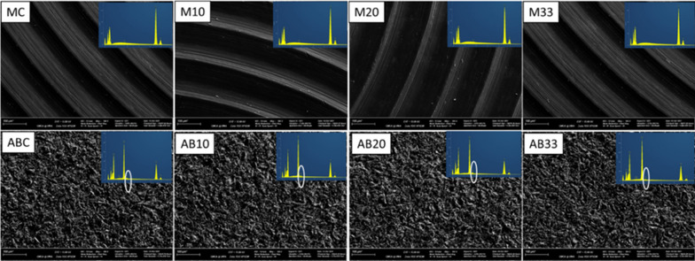
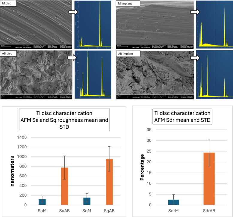
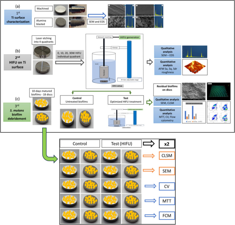
Methods: To optimise the HIFU parameters, four quadrants of a pair of Ti discs [machined (M) and alumina grit blasted (AB)] were marked using laser etching (MD Waterlase, US). HIFU beams, generated by a 254 kHz transducer and operated at intensities of 0 W, 10 W, 20 W, and 30 W, were applied to each quadrant for 2 min (min) in a water medium. The roughness of the treated surfaces was measured using Atomic Force Microscopy (AFM), and the surface composition was analyzed using Scanning Electron Microscope-Energy Dispersive Spectrometry (SEM-EDS). To investigate the biofilm debridement, 10-day-old S. mutans cultures were grown on 20 pairs of similar Ti discs, and then the optimized HIFU intensity of 20W was applied to five test pairs. Qualitative analyses were performed using a Dual Fluorescence/Reflection Confocal Laser Scanning Microscope (FRCLSM) and SEM imaging. Quantitative data on cell viability were collected using crystal violet (CV), (3-[4,5-dimethylthiazol-2-yl]-2,5 2,5-diphenyl tetrazolium bromide) (MTT), and flow cytometry (FCM) assays. Data from these test conditions were analyzed alongside cultures on biofilms that were untreated (control). Statistical data were calculated using ANOVA and appropriate t-tests for repeated measures.
Results: The surface roughness of AB Ti discs showed a highest and significant increase (p < 0.05) following HIFU exposure at 20 W through three roughness parameters (Sa, Sq, and Sdr), compared to the controls (1207 nm, 1455 nm, 62% compared to 842 nm, 1042 nm and 30% respectively). This optimized HIFU treatment not only significantly reduced the bacterial counts of the biofilms (76% of total bacteria from M discs, 59% on AB discs in FCM assays) but also created areas of complete biofilm removal in SEM images.
Conclusion: This study provides preliminary in vitro evidence that HIFU can remove bacterial biofilms. Further research is required to determine its feasibility as a potential non-surgical approach for the prevention and management of peri-implantitis.
期刊介绍:
The International Journal of Implant Dentistry is a peer-reviewed open access journal published under the SpringerOpen brand. The journal is dedicated to promoting the exchange and discussion of all research areas relevant to implant dentistry in the form of systematic literature or invited reviews, prospective and retrospective clinical studies, clinical case reports, basic laboratory and animal research, and articles on material research and engineering.
 求助内容:
求助内容: 应助结果提醒方式:


