{"title":"甲状腺癌患者心肌血流和心血管功能改变及相关治疗的评价。","authors":"Daniel Hueng-Yuan Shen, Hung-Pin Chan, Chin Hu, Wen-Hwa Wang, Yu-Li Chiu, Fu-Ren Tsai","doi":"10.21873/invivo.14092","DOIUrl":null,"url":null,"abstract":"<p><strong>Background/aim: </strong>This study aimed to explore changes in cardiovascular performance among patients with thyroid cancer following thyroidectomy, radioiodine therapy (RAIT) and thyroid-stimulating hormone suppression therapy (TST), with or without treatment with tyrosine kinase inhibitors (TKIs).</p><p><strong>Patients and methods: </strong>We enrolled 32 patients who underwent thyroidectomy and subsequent RAIT (except for one patient who underwent partial thyroidectomy only) and TST, with or without TKI therapy. We assessed myocardial perfusion using quantitative myocardial perfusion imaging (qMPI), myocardial blood flow (MBF), and coronary flow reserve (CFR) <i>via</i> dynamic single photon-emission computed tomography/computer tomography. We also analyzed data from laboratory tests or heart-related circulation markers, such as <i>N</i>-terminal pro B-type natriuretic peptide (NT-proBNP) to compare patient results, including correlations with different TKIs, RAIT, and TST.</p><p><strong>Results: </strong>qMPI indicated that the TKI-treated group (n=19) exhibited reduced MBF, CFR, and coronary flow capacity compared to the non-TKI-treated group. The right coronary artery (RCA) territory was notably affected by TKI treatment, particularly during the stress phase: RCA-stress blood flow: 2.4±0.6 <i>vs.</i> 1.9±0.7 ml/min/g; RCA-CFR: 2.3±0.8 <i>vs.</i> 2.93±0.9 for TKI-treated <i>vs.</i> non TKI-treated groups, respectively. This effect was more pronounced in the subgroup undergoing sequential anti-vascular endothelial growth factor TKI treatment (<i>e.g.</i> sorafenib and lenvatinib) (n=4) compared to non-TKI-treated controls. Additionally, TKI-treated patients showed a higher mean NT-ProBNP level (203±322.2 pg/ml) than non-TKI-treated patients (39.1±36 pg/ml). Abnormal MBF/CFR was also associated with higher cumulative RAIT dose and lower thyrotropin level.</p><p><strong>Conclusion: </strong>TKI therapy, especially anti-vascular endothelial growth factor TKI, can negatively affect myocardial perfusion and lead to coronary microcirculation dysfunction, reducing MBF/CFR during the stress phase. The link between cumulative RAIT dose, lower thyroid-stimulating hormone level, and abnormal MBF/CFR highlights the need for further research.</p>","PeriodicalId":13364,"journal":{"name":"In vivo","volume":"39 5","pages":"2919-2930"},"PeriodicalIF":1.8000,"publicationDate":"2025-09-01","publicationTypes":"Journal Article","fieldsOfStudy":null,"isOpenAccess":false,"openAccessPdf":"https://www.ncbi.nlm.nih.gov/pmc/articles/PMC12396053/pdf/","citationCount":"0","resultStr":"{\"title\":\"Evaluation of Altered Myocardial Blood Flow and Cardiovascular Function in Patients With Thyroid Cancer and Related Treatments.\",\"authors\":\"Daniel Hueng-Yuan Shen, Hung-Pin Chan, Chin Hu, Wen-Hwa Wang, Yu-Li Chiu, Fu-Ren Tsai\",\"doi\":\"10.21873/invivo.14092\",\"DOIUrl\":null,\"url\":null,\"abstract\":\"<p><strong>Background/aim: </strong>This study aimed to explore changes in cardiovascular performance among patients with thyroid cancer following thyroidectomy, radioiodine therapy (RAIT) and thyroid-stimulating hormone suppression therapy (TST), with or without treatment with tyrosine kinase inhibitors (TKIs).</p><p><strong>Patients and methods: </strong>We enrolled 32 patients who underwent thyroidectomy and subsequent RAIT (except for one patient who underwent partial thyroidectomy only) and TST, with or without TKI therapy. We assessed myocardial perfusion using quantitative myocardial perfusion imaging (qMPI), myocardial blood flow (MBF), and coronary flow reserve (CFR) <i>via</i> dynamic single photon-emission computed tomography/computer tomography. We also analyzed data from laboratory tests or heart-related circulation markers, such as <i>N</i>-terminal pro B-type natriuretic peptide (NT-proBNP) to compare patient results, including correlations with different TKIs, RAIT, and TST.</p><p><strong>Results: </strong>qMPI indicated that the TKI-treated group (n=19) exhibited reduced MBF, CFR, and coronary flow capacity compared to the non-TKI-treated group. The right coronary artery (RCA) territory was notably affected by TKI treatment, particularly during the stress phase: RCA-stress blood flow: 2.4±0.6 <i>vs.</i> 1.9±0.7 ml/min/g; RCA-CFR: 2.3±0.8 <i>vs.</i> 2.93±0.9 for TKI-treated <i>vs.</i> non TKI-treated groups, respectively. This effect was more pronounced in the subgroup undergoing sequential anti-vascular endothelial growth factor TKI treatment (<i>e.g.</i> sorafenib and lenvatinib) (n=4) compared to non-TKI-treated controls. Additionally, TKI-treated patients showed a higher mean NT-ProBNP level (203±322.2 pg/ml) than non-TKI-treated patients (39.1±36 pg/ml). Abnormal MBF/CFR was also associated with higher cumulative RAIT dose and lower thyrotropin level.</p><p><strong>Conclusion: </strong>TKI therapy, especially anti-vascular endothelial growth factor TKI, can negatively affect myocardial perfusion and lead to coronary microcirculation dysfunction, reducing MBF/CFR during the stress phase. The link between cumulative RAIT dose, lower thyroid-stimulating hormone level, and abnormal MBF/CFR highlights the need for further research.</p>\",\"PeriodicalId\":13364,\"journal\":{\"name\":\"In vivo\",\"volume\":\"39 5\",\"pages\":\"2919-2930\"},\"PeriodicalIF\":1.8000,\"publicationDate\":\"2025-09-01\",\"publicationTypes\":\"Journal Article\",\"fieldsOfStudy\":null,\"isOpenAccess\":false,\"openAccessPdf\":\"https://www.ncbi.nlm.nih.gov/pmc/articles/PMC12396053/pdf/\",\"citationCount\":\"0\",\"resultStr\":null,\"platform\":\"Semanticscholar\",\"paperid\":null,\"PeriodicalName\":\"In vivo\",\"FirstCategoryId\":\"3\",\"ListUrlMain\":\"https://doi.org/10.21873/invivo.14092\",\"RegionNum\":4,\"RegionCategory\":\"医学\",\"ArticlePicture\":[],\"TitleCN\":null,\"AbstractTextCN\":null,\"PMCID\":null,\"EPubDate\":\"\",\"PubModel\":\"\",\"JCR\":\"Q3\",\"JCRName\":\"MEDICINE, RESEARCH & EXPERIMENTAL\",\"Score\":null,\"Total\":0}","platform":"Semanticscholar","paperid":null,"PeriodicalName":"In vivo","FirstCategoryId":"3","ListUrlMain":"https://doi.org/10.21873/invivo.14092","RegionNum":4,"RegionCategory":"医学","ArticlePicture":[],"TitleCN":null,"AbstractTextCN":null,"PMCID":null,"EPubDate":"","PubModel":"","JCR":"Q3","JCRName":"MEDICINE, RESEARCH & EXPERIMENTAL","Score":null,"Total":0}
引用次数: 0
摘要
背景/目的:本研究旨在探讨甲状腺癌患者在甲状腺切除术、放射性碘治疗(RAIT)和促甲状腺激素抑制治疗(TST)后,接受或不接受酪氨酸激酶抑制剂(TKIs)治疗后心血管功能的变化。患者和方法:我们招募了32例接受甲状腺切除术和随后的RAIT(除了1例仅接受部分甲状腺切除术的患者)和TST的患者,有或没有TKI治疗。我们使用定量心肌灌注成像(qMPI)评估心肌灌注,心肌血流量(MBF)和冠状动脉血流储备(CFR)通过动态单光子发射计算机断层扫描/计算机断层扫描。我们还分析了实验室检测或心脏相关循环标志物的数据,如n端前b型利钠肽(NT-proBNP),以比较患者结果,包括与不同TKIs、RAIT和TST的相关性。结果:qMPI显示,与非tki治疗组相比,tki治疗组(n=19)表现出MBF、CFR和冠状动脉血流容量的降低。TKI治疗显著影响右冠状动脉(RCA)区域,特别是在应激期:RCA应激血流量:2.4±0.6 vs 1.9±0.7 ml/min/g;tki治疗组和非tki治疗组的RCA-CFR分别为2.3±0.8和2.93±0.9。与未接受TKI治疗的对照组相比,接受序贯抗血管内皮生长因子TKI治疗(如索拉非尼和lenvatinib)的亚组(n=4)的效果更为明显。此外,tki治疗患者的NT-ProBNP平均水平(203±322.2 pg/ml)高于未tki治疗患者(39.1±36 pg/ml)。异常的MBF/CFR也与较高的累积RAIT剂量和较低的促甲状腺激素水平相关。结论:TKI治疗,特别是抗血管内皮生长因子TKI治疗可影响心肌灌注,导致冠状动脉微循环功能障碍,降低应激期MBF/CFR。累积RAIT剂量、较低促甲状腺激素水平和异常MBF/CFR之间的联系强调了进一步研究的必要性。
Evaluation of Altered Myocardial Blood Flow and Cardiovascular Function in Patients With Thyroid Cancer and Related Treatments.
Background/aim: This study aimed to explore changes in cardiovascular performance among patients with thyroid cancer following thyroidectomy, radioiodine therapy (RAIT) and thyroid-stimulating hormone suppression therapy (TST), with or without treatment with tyrosine kinase inhibitors (TKIs).
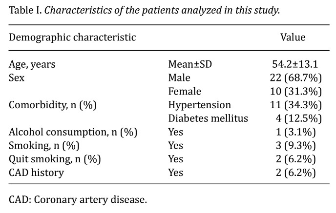
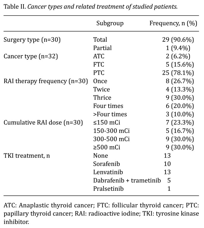
Patients and methods: We enrolled 32 patients who underwent thyroidectomy and subsequent RAIT (except for one patient who underwent partial thyroidectomy only) and TST, with or without TKI therapy. We assessed myocardial perfusion using quantitative myocardial perfusion imaging (qMPI), myocardial blood flow (MBF), and coronary flow reserve (CFR) via dynamic single photon-emission computed tomography/computer tomography. We also analyzed data from laboratory tests or heart-related circulation markers, such as N-terminal pro B-type natriuretic peptide (NT-proBNP) to compare patient results, including correlations with different TKIs, RAIT, and TST.
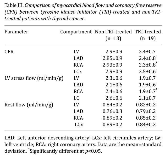
Results: qMPI indicated that the TKI-treated group (n=19) exhibited reduced MBF, CFR, and coronary flow capacity compared to the non-TKI-treated group. The right coronary artery (RCA) territory was notably affected by TKI treatment, particularly during the stress phase: RCA-stress blood flow: 2.4±0.6 vs. 1.9±0.7 ml/min/g; RCA-CFR: 2.3±0.8 vs. 2.93±0.9 for TKI-treated vs. non TKI-treated groups, respectively. This effect was more pronounced in the subgroup undergoing sequential anti-vascular endothelial growth factor TKI treatment (e.g. sorafenib and lenvatinib) (n=4) compared to non-TKI-treated controls. Additionally, TKI-treated patients showed a higher mean NT-ProBNP level (203±322.2 pg/ml) than non-TKI-treated patients (39.1±36 pg/ml). Abnormal MBF/CFR was also associated with higher cumulative RAIT dose and lower thyrotropin level.
Conclusion: TKI therapy, especially anti-vascular endothelial growth factor TKI, can negatively affect myocardial perfusion and lead to coronary microcirculation dysfunction, reducing MBF/CFR during the stress phase. The link between cumulative RAIT dose, lower thyroid-stimulating hormone level, and abnormal MBF/CFR highlights the need for further research.
期刊介绍:
IN VIVO is an international peer-reviewed journal designed to bring together original high quality works and reviews on experimental and clinical biomedical research within the frames of physiology, pathology and disease management.
The topics of IN VIVO include: 1. Experimental development and application of new diagnostic and therapeutic procedures; 2. Pharmacological and toxicological evaluation of new drugs, drug combinations and drug delivery systems; 3. Clinical trials; 4. Development and characterization of models of biomedical research; 5. Cancer diagnosis and treatment; 6. Immunotherapy and vaccines; 7. Radiotherapy, Imaging; 8. Tissue engineering, Regenerative medicine; 9. Carcinogenesis.

 求助内容:
求助内容: 应助结果提醒方式:


