{"title":"抑制巨噬细胞VEGFR1 TK信号可抑制子宫内膜异位症的发生。","authors":"Akiko Furue, Masako Honda, Kyoko Hattori, Erina Sato, Atsushi Yamashita, Mayuko Osada, Kanako Hosono, Mariko Kamata, Yoshiya Ito, Masabumi Shibuya, Kazuyoshi Kato, Hideki Amano","doi":"10.21873/invivo.14059","DOIUrl":null,"url":null,"abstract":"<p><strong>Background/aim: </strong>Endometriosis is characterized by the accumulation of immune cells in endometrial lesions and the peritoneal cavity. Macrophages contribute to the growth and neovascularization of endometriotic lesions. Vascular endothelial growth factor receptor-1 (VEGFR1) is involved in neovascularization, while peritoneal macrophages (PMs) play a critical role in endometriosis development and establishment. We examined the role of VEGFR1 signaling in PMs during endometriosis development using a murine model of ectopic endometrial transplantation.</p><p><strong>Materials and methods: </strong>Endometrial fragments from female wild-type (WT) or VEGFR1 tyrosine kinase-deficient (TK<sup>-/-</sup>) donor mice were implanted into the peritoneal walls of recipient mice, either in a WT→WT or TK<sup>-/-</sup>→TK<sup>-/-</sup> combination. On day 14 after endometrial transplantation, the implant size, neovascular growth-promoting factors, macrophage accumulation in the implants and peritoneal cavity, and cytokine production were assessed. PMs from WT or TK<sup>-/-</sup> mice were transferred into the peritoneal cavity of WT→WT mice and their effects were assessed.</p><p><strong>Results: </strong>Compared to WT→WT mice, TK<sup>-/-</sup>→TK<sup>-/-</sup> mice exhibited smaller implant sizes and reduced neovascularization, including angiogenesis and lymphangiogenesis. This was correlated with an increase in pro-inflammatory (M1) and a decrease in alternative (M2) large peritoneal macrophages (LPMs) within the peritoneal cavity. Transfer of TK<sup>-/-</sup>-PMs into the peritoneal cavity of WT→WT mice reduced endometriosis development and macrophage accumulation. This led to increased expression of M1 macrophage genes and decreased expression of M2 phenotype genes, compared to WT-PMs transfer. PMs from TK<sup>-/-</sup> mice exhibited increased M1-related and decreased M2-related gene expression.</p><p><strong>Conclusion: </strong>Deletion of VEGFR1 TK signaling in PMs suppressed endometriosis progression and neovascularization by increasing M1 LPMs. Specific inactivation of VEGFR1 TK signaling may represent a potential therapeutic target for the management of endometriosis.</p>","PeriodicalId":13364,"journal":{"name":"In vivo","volume":"39 5","pages":"2584-2598"},"PeriodicalIF":1.8000,"publicationDate":"2025-09-01","publicationTypes":"Journal Article","fieldsOfStudy":null,"isOpenAccess":false,"openAccessPdf":"https://www.ncbi.nlm.nih.gov/pmc/articles/PMC12396080/pdf/","citationCount":"0","resultStr":"{\"title\":\"Inhibition of VEGFR1 TK Signaling in Peritoneal Macrophages Suppresses Endometriosis Development.\",\"authors\":\"Akiko Furue, Masako Honda, Kyoko Hattori, Erina Sato, Atsushi Yamashita, Mayuko Osada, Kanako Hosono, Mariko Kamata, Yoshiya Ito, Masabumi Shibuya, Kazuyoshi Kato, Hideki Amano\",\"doi\":\"10.21873/invivo.14059\",\"DOIUrl\":null,\"url\":null,\"abstract\":\"<p><strong>Background/aim: </strong>Endometriosis is characterized by the accumulation of immune cells in endometrial lesions and the peritoneal cavity. Macrophages contribute to the growth and neovascularization of endometriotic lesions. Vascular endothelial growth factor receptor-1 (VEGFR1) is involved in neovascularization, while peritoneal macrophages (PMs) play a critical role in endometriosis development and establishment. We examined the role of VEGFR1 signaling in PMs during endometriosis development using a murine model of ectopic endometrial transplantation.</p><p><strong>Materials and methods: </strong>Endometrial fragments from female wild-type (WT) or VEGFR1 tyrosine kinase-deficient (TK<sup>-/-</sup>) donor mice were implanted into the peritoneal walls of recipient mice, either in a WT→WT or TK<sup>-/-</sup>→TK<sup>-/-</sup> combination. On day 14 after endometrial transplantation, the implant size, neovascular growth-promoting factors, macrophage accumulation in the implants and peritoneal cavity, and cytokine production were assessed. PMs from WT or TK<sup>-/-</sup> mice were transferred into the peritoneal cavity of WT→WT mice and their effects were assessed.</p><p><strong>Results: </strong>Compared to WT→WT mice, TK<sup>-/-</sup>→TK<sup>-/-</sup> mice exhibited smaller implant sizes and reduced neovascularization, including angiogenesis and lymphangiogenesis. This was correlated with an increase in pro-inflammatory (M1) and a decrease in alternative (M2) large peritoneal macrophages (LPMs) within the peritoneal cavity. Transfer of TK<sup>-/-</sup>-PMs into the peritoneal cavity of WT→WT mice reduced endometriosis development and macrophage accumulation. This led to increased expression of M1 macrophage genes and decreased expression of M2 phenotype genes, compared to WT-PMs transfer. PMs from TK<sup>-/-</sup> mice exhibited increased M1-related and decreased M2-related gene expression.</p><p><strong>Conclusion: </strong>Deletion of VEGFR1 TK signaling in PMs suppressed endometriosis progression and neovascularization by increasing M1 LPMs. Specific inactivation of VEGFR1 TK signaling may represent a potential therapeutic target for the management of endometriosis.</p>\",\"PeriodicalId\":13364,\"journal\":{\"name\":\"In vivo\",\"volume\":\"39 5\",\"pages\":\"2584-2598\"},\"PeriodicalIF\":1.8000,\"publicationDate\":\"2025-09-01\",\"publicationTypes\":\"Journal Article\",\"fieldsOfStudy\":null,\"isOpenAccess\":false,\"openAccessPdf\":\"https://www.ncbi.nlm.nih.gov/pmc/articles/PMC12396080/pdf/\",\"citationCount\":\"0\",\"resultStr\":null,\"platform\":\"Semanticscholar\",\"paperid\":null,\"PeriodicalName\":\"In vivo\",\"FirstCategoryId\":\"3\",\"ListUrlMain\":\"https://doi.org/10.21873/invivo.14059\",\"RegionNum\":4,\"RegionCategory\":\"医学\",\"ArticlePicture\":[],\"TitleCN\":null,\"AbstractTextCN\":null,\"PMCID\":null,\"EPubDate\":\"\",\"PubModel\":\"\",\"JCR\":\"Q3\",\"JCRName\":\"MEDICINE, RESEARCH & EXPERIMENTAL\",\"Score\":null,\"Total\":0}","platform":"Semanticscholar","paperid":null,"PeriodicalName":"In vivo","FirstCategoryId":"3","ListUrlMain":"https://doi.org/10.21873/invivo.14059","RegionNum":4,"RegionCategory":"医学","ArticlePicture":[],"TitleCN":null,"AbstractTextCN":null,"PMCID":null,"EPubDate":"","PubModel":"","JCR":"Q3","JCRName":"MEDICINE, RESEARCH & EXPERIMENTAL","Score":null,"Total":0}
Inhibition of VEGFR1 TK Signaling in Peritoneal Macrophages Suppresses Endometriosis Development.
Background/aim: Endometriosis is characterized by the accumulation of immune cells in endometrial lesions and the peritoneal cavity. Macrophages contribute to the growth and neovascularization of endometriotic lesions. Vascular endothelial growth factor receptor-1 (VEGFR1) is involved in neovascularization, while peritoneal macrophages (PMs) play a critical role in endometriosis development and establishment. We examined the role of VEGFR1 signaling in PMs during endometriosis development using a murine model of ectopic endometrial transplantation.
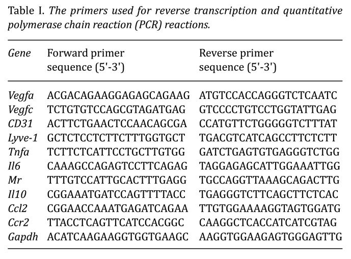
Materials and methods: Endometrial fragments from female wild-type (WT) or VEGFR1 tyrosine kinase-deficient (TK-/-) donor mice were implanted into the peritoneal walls of recipient mice, either in a WT→WT or TK-/-→TK-/- combination. On day 14 after endometrial transplantation, the implant size, neovascular growth-promoting factors, macrophage accumulation in the implants and peritoneal cavity, and cytokine production were assessed. PMs from WT or TK-/- mice were transferred into the peritoneal cavity of WT→WT mice and their effects were assessed.
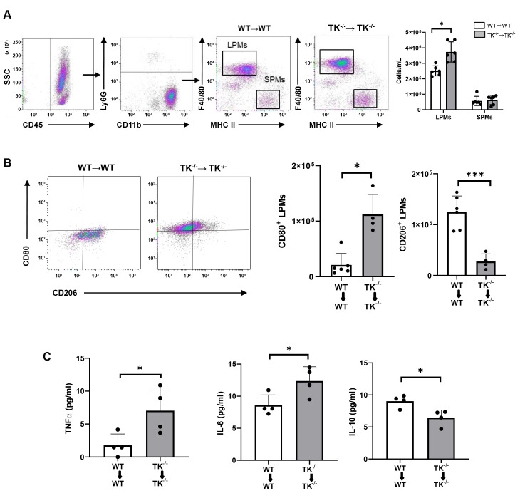
Results: Compared to WT→WT mice, TK-/-→TK-/- mice exhibited smaller implant sizes and reduced neovascularization, including angiogenesis and lymphangiogenesis. This was correlated with an increase in pro-inflammatory (M1) and a decrease in alternative (M2) large peritoneal macrophages (LPMs) within the peritoneal cavity. Transfer of TK-/--PMs into the peritoneal cavity of WT→WT mice reduced endometriosis development and macrophage accumulation. This led to increased expression of M1 macrophage genes and decreased expression of M2 phenotype genes, compared to WT-PMs transfer. PMs from TK-/- mice exhibited increased M1-related and decreased M2-related gene expression.
Conclusion: Deletion of VEGFR1 TK signaling in PMs suppressed endometriosis progression and neovascularization by increasing M1 LPMs. Specific inactivation of VEGFR1 TK signaling may represent a potential therapeutic target for the management of endometriosis.
期刊介绍:
IN VIVO is an international peer-reviewed journal designed to bring together original high quality works and reviews on experimental and clinical biomedical research within the frames of physiology, pathology and disease management.
The topics of IN VIVO include: 1. Experimental development and application of new diagnostic and therapeutic procedures; 2. Pharmacological and toxicological evaluation of new drugs, drug combinations and drug delivery systems; 3. Clinical trials; 4. Development and characterization of models of biomedical research; 5. Cancer diagnosis and treatment; 6. Immunotherapy and vaccines; 7. Radiotherapy, Imaging; 8. Tissue engineering, Regenerative medicine; 9. Carcinogenesis.


 求助内容:
求助内容: 应助结果提醒方式:


