{"title":"再次检查浅表性血管粘液瘤。","authors":"Yuki Shinohara, Yoshiro Chijiiwa, Jun Nishio","doi":"10.21873/invivo.14053","DOIUrl":null,"url":null,"abstract":"<p><p>Superficial angiomyxoma (SAM) is a rare benign mesenchymal tumor of uncertain differentiation that primarily occurs in the trunk, lower extremities and head and neck of middle-aged adults. It typically presents as a slow-growing, painless polypoid or papulonodular lesion. Ultrasonography shows an ovoid, well-defined mass with a homogeneous, hypoechoic echotexture. Color Doppler examination may demonstrate prominent hypervascularity. Computed tomography reveals a round to oval hypodense mass. On magnetic resonance imaging, SAM usually exhibits low to intermediate signal intensity on T1-weighted sequences and high signal intensity on T2-weighted sequences. Heterogenous enhancement is seen after intravenous contrast administration. Histologically, the lesion is composed of bland spindle-shaped and stellate-shaped cells in an abundant myxoid stroma with numerous small blood vessels. By immunohistochemistry, the tumor cells are positive for CD34 and show variable staining for smooth muscle actin and S-100 protein. Loss of protein kinase cAMP-dependent type I regulatory subunit alpha (PRKAR1A) expression is observed in a significant subset of cases. Complete surgical excision is the treatment of choice for SAM. This review provides an updated overview of the clinicopathological, radiological and genomic features of SAM and discusses the differential diagnosis of this peculiar tumor.</p>","PeriodicalId":13364,"journal":{"name":"In vivo","volume":"39 5","pages":"2505-2511"},"PeriodicalIF":1.8000,"publicationDate":"2025-09-01","publicationTypes":"Journal Article","fieldsOfStudy":null,"isOpenAccess":false,"openAccessPdf":"https://www.ncbi.nlm.nih.gov/pmc/articles/PMC12396051/pdf/","citationCount":"0","resultStr":"{\"title\":\"Superficial Angiomyxoma Revisited.\",\"authors\":\"Yuki Shinohara, Yoshiro Chijiiwa, Jun Nishio\",\"doi\":\"10.21873/invivo.14053\",\"DOIUrl\":null,\"url\":null,\"abstract\":\"<p><p>Superficial angiomyxoma (SAM) is a rare benign mesenchymal tumor of uncertain differentiation that primarily occurs in the trunk, lower extremities and head and neck of middle-aged adults. It typically presents as a slow-growing, painless polypoid or papulonodular lesion. Ultrasonography shows an ovoid, well-defined mass with a homogeneous, hypoechoic echotexture. Color Doppler examination may demonstrate prominent hypervascularity. Computed tomography reveals a round to oval hypodense mass. On magnetic resonance imaging, SAM usually exhibits low to intermediate signal intensity on T1-weighted sequences and high signal intensity on T2-weighted sequences. Heterogenous enhancement is seen after intravenous contrast administration. Histologically, the lesion is composed of bland spindle-shaped and stellate-shaped cells in an abundant myxoid stroma with numerous small blood vessels. By immunohistochemistry, the tumor cells are positive for CD34 and show variable staining for smooth muscle actin and S-100 protein. Loss of protein kinase cAMP-dependent type I regulatory subunit alpha (PRKAR1A) expression is observed in a significant subset of cases. Complete surgical excision is the treatment of choice for SAM. This review provides an updated overview of the clinicopathological, radiological and genomic features of SAM and discusses the differential diagnosis of this peculiar tumor.</p>\",\"PeriodicalId\":13364,\"journal\":{\"name\":\"In vivo\",\"volume\":\"39 5\",\"pages\":\"2505-2511\"},\"PeriodicalIF\":1.8000,\"publicationDate\":\"2025-09-01\",\"publicationTypes\":\"Journal Article\",\"fieldsOfStudy\":null,\"isOpenAccess\":false,\"openAccessPdf\":\"https://www.ncbi.nlm.nih.gov/pmc/articles/PMC12396051/pdf/\",\"citationCount\":\"0\",\"resultStr\":null,\"platform\":\"Semanticscholar\",\"paperid\":null,\"PeriodicalName\":\"In vivo\",\"FirstCategoryId\":\"3\",\"ListUrlMain\":\"https://doi.org/10.21873/invivo.14053\",\"RegionNum\":4,\"RegionCategory\":\"医学\",\"ArticlePicture\":[],\"TitleCN\":null,\"AbstractTextCN\":null,\"PMCID\":null,\"EPubDate\":\"\",\"PubModel\":\"\",\"JCR\":\"Q3\",\"JCRName\":\"MEDICINE, RESEARCH & EXPERIMENTAL\",\"Score\":null,\"Total\":0}","platform":"Semanticscholar","paperid":null,"PeriodicalName":"In vivo","FirstCategoryId":"3","ListUrlMain":"https://doi.org/10.21873/invivo.14053","RegionNum":4,"RegionCategory":"医学","ArticlePicture":[],"TitleCN":null,"AbstractTextCN":null,"PMCID":null,"EPubDate":"","PubModel":"","JCR":"Q3","JCRName":"MEDICINE, RESEARCH & EXPERIMENTAL","Score":null,"Total":0}
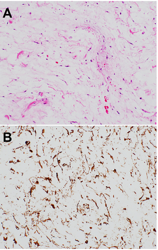
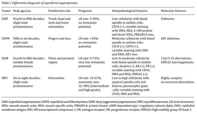
Superficial angiomyxoma (SAM) is a rare benign mesenchymal tumor of uncertain differentiation that primarily occurs in the trunk, lower extremities and head and neck of middle-aged adults. It typically presents as a slow-growing, painless polypoid or papulonodular lesion. Ultrasonography shows an ovoid, well-defined mass with a homogeneous, hypoechoic echotexture. Color Doppler examination may demonstrate prominent hypervascularity. Computed tomography reveals a round to oval hypodense mass. On magnetic resonance imaging, SAM usually exhibits low to intermediate signal intensity on T1-weighted sequences and high signal intensity on T2-weighted sequences. Heterogenous enhancement is seen after intravenous contrast administration. Histologically, the lesion is composed of bland spindle-shaped and stellate-shaped cells in an abundant myxoid stroma with numerous small blood vessels. By immunohistochemistry, the tumor cells are positive for CD34 and show variable staining for smooth muscle actin and S-100 protein. Loss of protein kinase cAMP-dependent type I regulatory subunit alpha (PRKAR1A) expression is observed in a significant subset of cases. Complete surgical excision is the treatment of choice for SAM. This review provides an updated overview of the clinicopathological, radiological and genomic features of SAM and discusses the differential diagnosis of this peculiar tumor.
期刊介绍:
IN VIVO is an international peer-reviewed journal designed to bring together original high quality works and reviews on experimental and clinical biomedical research within the frames of physiology, pathology and disease management.
The topics of IN VIVO include: 1. Experimental development and application of new diagnostic and therapeutic procedures; 2. Pharmacological and toxicological evaluation of new drugs, drug combinations and drug delivery systems; 3. Clinical trials; 4. Development and characterization of models of biomedical research; 5. Cancer diagnosis and treatment; 6. Immunotherapy and vaccines; 7. Radiotherapy, Imaging; 8. Tissue engineering, Regenerative medicine; 9. Carcinogenesis.

 求助内容:
求助内容: 应助结果提醒方式:


