Georgia Vairaktari, Efstathia Vairaktari, Alexander Schramm, Spyridoula Derka, Veronika Papakosta, Spyridon Stavrianos, Aikaterini Bini, Andreas Sakkas, Maria Kouri, Antonis Vylliotis, Andreas Lazaris, Marcel Ebeling
{"title":"通过p53和p63联合表达分析对皮肤癌患者进行分层并指导治疗决策。","authors":"Georgia Vairaktari, Efstathia Vairaktari, Alexander Schramm, Spyridoula Derka, Veronika Papakosta, Spyridon Stavrianos, Aikaterini Bini, Andreas Sakkas, Maria Kouri, Antonis Vylliotis, Andreas Lazaris, Marcel Ebeling","doi":"10.21873/invivo.14060","DOIUrl":null,"url":null,"abstract":"<p><strong>Background/aim: </strong>Skin cancer, particularly non-melanocytic types like squamous and basal cell carcinoma, remains a growing concern. The tumor suppressor proteins p53 and p63 play key roles in skin carcinogenesis. This study aimed to assess the differential expression of p53 and p63 in various stages of chemically-induced skin cancer.</p><p><strong>Materials and methods: </strong>FVB/N mice, aged 44 weeks, were randomly assigned into three groups: a control group (n=8) and two experimental groups (Group A: n=16, Group B: n=16). The study employed a two-stage carcinogenesis procedure, which involved an initial application of 97.4 nmol DMBA to shaved skin on the back, followed by applications of 32.4 nmol TPA after thirteen weeks for Group A and after twenty weeks for Group B. The control group did not receive any treatment. Skin lesions were monitored, and tissue samples were collected for histological and immunohistochemical analysis.</p><p><strong>Results: </strong>p53 expression was significantly elevated in precancerous and benign tumors compared to normal histology (47.6% and 47.8% <i>vs.</i> 18.8%, respectively; <i>p</i><0.05), but not in malignant tumors. Mean p53 expression was significantly higher in both experimental groups compared to controls (group A: 42.1%, group B: 47.1%; <i>p</i><0.001). Conversely, p63 expression remained generally low across all stages, with slightly higher levels in malignant lesions. The difference in expression between p53 and p63 was significant in precancerous and benign lesions (<i>p</i><0.001). No significant differences in expression were found between the two experimental groups.</p><p><strong>Conclusion: </strong>Distinct expression patterns of p53 and p63 suggest stage-specific roles in skin carcinogenesis. Elevated p53 in early lesions supports its tumor-suppressive function, while p63 may contribute to tumor maintenance in advanced stages. These findings support the utility of p53 and p63 as biomarkers for diagnosis and prognosis in skin cancer, and potential targets for future therapies.</p>","PeriodicalId":13364,"journal":{"name":"In vivo","volume":"39 5","pages":"2599-2608"},"PeriodicalIF":1.8000,"publicationDate":"2025-09-01","publicationTypes":"Journal Article","fieldsOfStudy":null,"isOpenAccess":false,"openAccessPdf":"https://www.ncbi.nlm.nih.gov/pmc/articles/PMC12396083/pdf/","citationCount":"0","resultStr":"{\"title\":\"Stratifying Skin Cancer Patients and Guiding Treatment Decisions Through Combined p53 and p63 Expression Analysis.\",\"authors\":\"Georgia Vairaktari, Efstathia Vairaktari, Alexander Schramm, Spyridoula Derka, Veronika Papakosta, Spyridon Stavrianos, Aikaterini Bini, Andreas Sakkas, Maria Kouri, Antonis Vylliotis, Andreas Lazaris, Marcel Ebeling\",\"doi\":\"10.21873/invivo.14060\",\"DOIUrl\":null,\"url\":null,\"abstract\":\"<p><strong>Background/aim: </strong>Skin cancer, particularly non-melanocytic types like squamous and basal cell carcinoma, remains a growing concern. The tumor suppressor proteins p53 and p63 play key roles in skin carcinogenesis. This study aimed to assess the differential expression of p53 and p63 in various stages of chemically-induced skin cancer.</p><p><strong>Materials and methods: </strong>FVB/N mice, aged 44 weeks, were randomly assigned into three groups: a control group (n=8) and two experimental groups (Group A: n=16, Group B: n=16). The study employed a two-stage carcinogenesis procedure, which involved an initial application of 97.4 nmol DMBA to shaved skin on the back, followed by applications of 32.4 nmol TPA after thirteen weeks for Group A and after twenty weeks for Group B. The control group did not receive any treatment. Skin lesions were monitored, and tissue samples were collected for histological and immunohistochemical analysis.</p><p><strong>Results: </strong>p53 expression was significantly elevated in precancerous and benign tumors compared to normal histology (47.6% and 47.8% <i>vs.</i> 18.8%, respectively; <i>p</i><0.05), but not in malignant tumors. Mean p53 expression was significantly higher in both experimental groups compared to controls (group A: 42.1%, group B: 47.1%; <i>p</i><0.001). Conversely, p63 expression remained generally low across all stages, with slightly higher levels in malignant lesions. The difference in expression between p53 and p63 was significant in precancerous and benign lesions (<i>p</i><0.001). No significant differences in expression were found between the two experimental groups.</p><p><strong>Conclusion: </strong>Distinct expression patterns of p53 and p63 suggest stage-specific roles in skin carcinogenesis. Elevated p53 in early lesions supports its tumor-suppressive function, while p63 may contribute to tumor maintenance in advanced stages. These findings support the utility of p53 and p63 as biomarkers for diagnosis and prognosis in skin cancer, and potential targets for future therapies.</p>\",\"PeriodicalId\":13364,\"journal\":{\"name\":\"In vivo\",\"volume\":\"39 5\",\"pages\":\"2599-2608\"},\"PeriodicalIF\":1.8000,\"publicationDate\":\"2025-09-01\",\"publicationTypes\":\"Journal Article\",\"fieldsOfStudy\":null,\"isOpenAccess\":false,\"openAccessPdf\":\"https://www.ncbi.nlm.nih.gov/pmc/articles/PMC12396083/pdf/\",\"citationCount\":\"0\",\"resultStr\":null,\"platform\":\"Semanticscholar\",\"paperid\":null,\"PeriodicalName\":\"In vivo\",\"FirstCategoryId\":\"3\",\"ListUrlMain\":\"https://doi.org/10.21873/invivo.14060\",\"RegionNum\":4,\"RegionCategory\":\"医学\",\"ArticlePicture\":[],\"TitleCN\":null,\"AbstractTextCN\":null,\"PMCID\":null,\"EPubDate\":\"\",\"PubModel\":\"\",\"JCR\":\"Q3\",\"JCRName\":\"MEDICINE, RESEARCH & EXPERIMENTAL\",\"Score\":null,\"Total\":0}","platform":"Semanticscholar","paperid":null,"PeriodicalName":"In vivo","FirstCategoryId":"3","ListUrlMain":"https://doi.org/10.21873/invivo.14060","RegionNum":4,"RegionCategory":"医学","ArticlePicture":[],"TitleCN":null,"AbstractTextCN":null,"PMCID":null,"EPubDate":"","PubModel":"","JCR":"Q3","JCRName":"MEDICINE, RESEARCH & EXPERIMENTAL","Score":null,"Total":0}
Stratifying Skin Cancer Patients and Guiding Treatment Decisions Through Combined p53 and p63 Expression Analysis.
Background/aim: Skin cancer, particularly non-melanocytic types like squamous and basal cell carcinoma, remains a growing concern. The tumor suppressor proteins p53 and p63 play key roles in skin carcinogenesis. This study aimed to assess the differential expression of p53 and p63 in various stages of chemically-induced skin cancer.
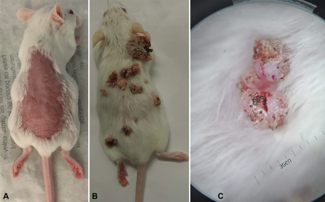
Materials and methods: FVB/N mice, aged 44 weeks, were randomly assigned into three groups: a control group (n=8) and two experimental groups (Group A: n=16, Group B: n=16). The study employed a two-stage carcinogenesis procedure, which involved an initial application of 97.4 nmol DMBA to shaved skin on the back, followed by applications of 32.4 nmol TPA after thirteen weeks for Group A and after twenty weeks for Group B. The control group did not receive any treatment. Skin lesions were monitored, and tissue samples were collected for histological and immunohistochemical analysis.
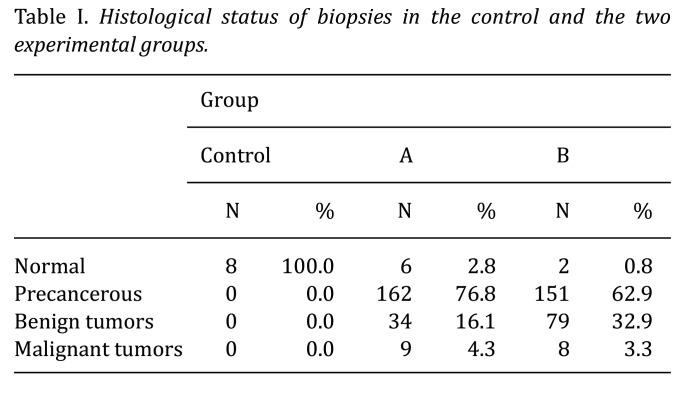
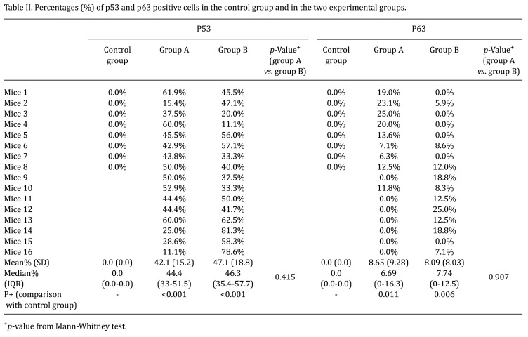
Results: p53 expression was significantly elevated in precancerous and benign tumors compared to normal histology (47.6% and 47.8% vs. 18.8%, respectively; p<0.05), but not in malignant tumors. Mean p53 expression was significantly higher in both experimental groups compared to controls (group A: 42.1%, group B: 47.1%; p<0.001). Conversely, p63 expression remained generally low across all stages, with slightly higher levels in malignant lesions. The difference in expression between p53 and p63 was significant in precancerous and benign lesions (p<0.001). No significant differences in expression were found between the two experimental groups.
Conclusion: Distinct expression patterns of p53 and p63 suggest stage-specific roles in skin carcinogenesis. Elevated p53 in early lesions supports its tumor-suppressive function, while p63 may contribute to tumor maintenance in advanced stages. These findings support the utility of p53 and p63 as biomarkers for diagnosis and prognosis in skin cancer, and potential targets for future therapies.
期刊介绍:
IN VIVO is an international peer-reviewed journal designed to bring together original high quality works and reviews on experimental and clinical biomedical research within the frames of physiology, pathology and disease management.
The topics of IN VIVO include: 1. Experimental development and application of new diagnostic and therapeutic procedures; 2. Pharmacological and toxicological evaluation of new drugs, drug combinations and drug delivery systems; 3. Clinical trials; 4. Development and characterization of models of biomedical research; 5. Cancer diagnosis and treatment; 6. Immunotherapy and vaccines; 7. Radiotherapy, Imaging; 8. Tissue engineering, Regenerative medicine; 9. Carcinogenesis.

 求助内容:
求助内容: 应助结果提醒方式:


