Jonas Müller, Gilles Dietrich, Igor Gossuin, Marc Prod'homme, Salah Dine Qanadli, Elyazid Mouhsine
{"title":"经皮跖骨远端截骨术矫正严重拇外翻畸形。","authors":"Jonas Müller, Gilles Dietrich, Igor Gossuin, Marc Prod'homme, Salah Dine Qanadli, Elyazid Mouhsine","doi":"10.1177/24730114251363448","DOIUrl":null,"url":null,"abstract":"<p><strong>Background: </strong>Severe hallux valgus (HV) presents a significant surgical challenge. Traditional methods are being questioned because of their invasiveness, whereas recent minimally invasive techniques raise concerns because of their associated complications. This study evaluates the percutaneous distal metatarsal osteotomy technique, generally found to be effective for mild-to-moderate cases, and tests the hypothesis that it provides effective clinical and radiologic correction for severe deformities.</p><p><strong>Methods: </strong>This retrospective study analyzed 116 feet that underwent percutaneous distal metatarsal transverse osteotomy with lateral soft tissue release and provisional Kirschner wire fixation, with a mean follow-up of 27.1 months, limited to severe cases (hallux valgus angle [HVA] > 40 degrees). Radiologic assessments included preoperative and postoperative measurements of HVA, intermetatarsal angle (IMA), distal metatarsal articular angle, sesamoid position, first metatarsophalangeal (MTPI) joint congruency, metatarsal length, and sagittal position. Clinical evaluations used the AOFAS scale, documenting the recurrence rate, the nature of complications, reoperations, and the association between them. Patient satisfaction was assessed through self-reported evaluations.</p><p><strong>Results: </strong>Significant improvements were noted for HVA (median correction from 43.1 to 14.6 degrees) and IMA (median correction from 17.2 to 8.5 degrees). The metatarsal was shortened by 5.4 mm. There was a notable reduction in the degree of sesamoid displacement and MTPI congruency. Sagittal position remained unchanged in 85.3%. The median AOFAS score improved from 44.0 to 90.5, well above the clinically significant improvement threshold, and 87.9% of patients were satisfied or very satisfied. We recorded no major complications and minor complications at a rate of 35.3%. Reoperation rate was 14.7%, primarily due to exostoses. Significant associations were found between postoperative sesamoid position and clinical outcome, and between reoperation rate, exostosis, and MTPI congruency, emphasizing the importance of correcting these parameters. Recurrence rate was 6%. Patient satisfaction was associated with reoperation and complications, but not with radiologic parameters.</p><p><strong>Conclusion: </strong>Percutaneous distal metatarsal osteotomy achieved substantial correction of severe hallux valgus with significant improvements in angular measurements, high patient satisfaction (87.9%), and no major complications. Although the technique shows promise as a less invasive alternative with comparable radiographic outcomes, the 14.7% reoperation rate (primarily for exostoses) and 6% recurrence rate must be considered. Prospective comparative studies are needed to establish its role relative to other surgical approaches for severe deformities.<b>Level of Evidence:</b> Level IV, retrospective case series.</p>","PeriodicalId":12429,"journal":{"name":"Foot & Ankle Orthopaedics","volume":"10 3","pages":"24730114251363448"},"PeriodicalIF":0.0000,"publicationDate":"2025-08-22","publicationTypes":"Journal Article","fieldsOfStudy":null,"isOpenAccess":false,"openAccessPdf":"https://www.ncbi.nlm.nih.gov/pmc/articles/PMC12374112/pdf/","citationCount":"0","resultStr":"{\"title\":\"Correction of Severe Hallux Valgus Deformity Using a Percutaneous Metatarsal Distal Osteotomy.\",\"authors\":\"Jonas Müller, Gilles Dietrich, Igor Gossuin, Marc Prod'homme, Salah Dine Qanadli, Elyazid Mouhsine\",\"doi\":\"10.1177/24730114251363448\",\"DOIUrl\":null,\"url\":null,\"abstract\":\"<p><strong>Background: </strong>Severe hallux valgus (HV) presents a significant surgical challenge. Traditional methods are being questioned because of their invasiveness, whereas recent minimally invasive techniques raise concerns because of their associated complications. This study evaluates the percutaneous distal metatarsal osteotomy technique, generally found to be effective for mild-to-moderate cases, and tests the hypothesis that it provides effective clinical and radiologic correction for severe deformities.</p><p><strong>Methods: </strong>This retrospective study analyzed 116 feet that underwent percutaneous distal metatarsal transverse osteotomy with lateral soft tissue release and provisional Kirschner wire fixation, with a mean follow-up of 27.1 months, limited to severe cases (hallux valgus angle [HVA] > 40 degrees). Radiologic assessments included preoperative and postoperative measurements of HVA, intermetatarsal angle (IMA), distal metatarsal articular angle, sesamoid position, first metatarsophalangeal (MTPI) joint congruency, metatarsal length, and sagittal position. Clinical evaluations used the AOFAS scale, documenting the recurrence rate, the nature of complications, reoperations, and the association between them. Patient satisfaction was assessed through self-reported evaluations.</p><p><strong>Results: </strong>Significant improvements were noted for HVA (median correction from 43.1 to 14.6 degrees) and IMA (median correction from 17.2 to 8.5 degrees). The metatarsal was shortened by 5.4 mm. There was a notable reduction in the degree of sesamoid displacement and MTPI congruency. Sagittal position remained unchanged in 85.3%. The median AOFAS score improved from 44.0 to 90.5, well above the clinically significant improvement threshold, and 87.9% of patients were satisfied or very satisfied. We recorded no major complications and minor complications at a rate of 35.3%. Reoperation rate was 14.7%, primarily due to exostoses. Significant associations were found between postoperative sesamoid position and clinical outcome, and between reoperation rate, exostosis, and MTPI congruency, emphasizing the importance of correcting these parameters. Recurrence rate was 6%. Patient satisfaction was associated with reoperation and complications, but not with radiologic parameters.</p><p><strong>Conclusion: </strong>Percutaneous distal metatarsal osteotomy achieved substantial correction of severe hallux valgus with significant improvements in angular measurements, high patient satisfaction (87.9%), and no major complications. Although the technique shows promise as a less invasive alternative with comparable radiographic outcomes, the 14.7% reoperation rate (primarily for exostoses) and 6% recurrence rate must be considered. Prospective comparative studies are needed to establish its role relative to other surgical approaches for severe deformities.<b>Level of Evidence:</b> Level IV, retrospective case series.</p>\",\"PeriodicalId\":12429,\"journal\":{\"name\":\"Foot & Ankle Orthopaedics\",\"volume\":\"10 3\",\"pages\":\"24730114251363448\"},\"PeriodicalIF\":0.0000,\"publicationDate\":\"2025-08-22\",\"publicationTypes\":\"Journal Article\",\"fieldsOfStudy\":null,\"isOpenAccess\":false,\"openAccessPdf\":\"https://www.ncbi.nlm.nih.gov/pmc/articles/PMC12374112/pdf/\",\"citationCount\":\"0\",\"resultStr\":null,\"platform\":\"Semanticscholar\",\"paperid\":null,\"PeriodicalName\":\"Foot & Ankle Orthopaedics\",\"FirstCategoryId\":\"1085\",\"ListUrlMain\":\"https://doi.org/10.1177/24730114251363448\",\"RegionNum\":0,\"RegionCategory\":null,\"ArticlePicture\":[],\"TitleCN\":null,\"AbstractTextCN\":null,\"PMCID\":null,\"EPubDate\":\"2025/7/1 0:00:00\",\"PubModel\":\"eCollection\",\"JCR\":\"\",\"JCRName\":\"\",\"Score\":null,\"Total\":0}","platform":"Semanticscholar","paperid":null,"PeriodicalName":"Foot & Ankle Orthopaedics","FirstCategoryId":"1085","ListUrlMain":"https://doi.org/10.1177/24730114251363448","RegionNum":0,"RegionCategory":null,"ArticlePicture":[],"TitleCN":null,"AbstractTextCN":null,"PMCID":null,"EPubDate":"2025/7/1 0:00:00","PubModel":"eCollection","JCR":"","JCRName":"","Score":null,"Total":0}
Correction of Severe Hallux Valgus Deformity Using a Percutaneous Metatarsal Distal Osteotomy.
Background: Severe hallux valgus (HV) presents a significant surgical challenge. Traditional methods are being questioned because of their invasiveness, whereas recent minimally invasive techniques raise concerns because of their associated complications. This study evaluates the percutaneous distal metatarsal osteotomy technique, generally found to be effective for mild-to-moderate cases, and tests the hypothesis that it provides effective clinical and radiologic correction for severe deformities.
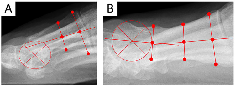
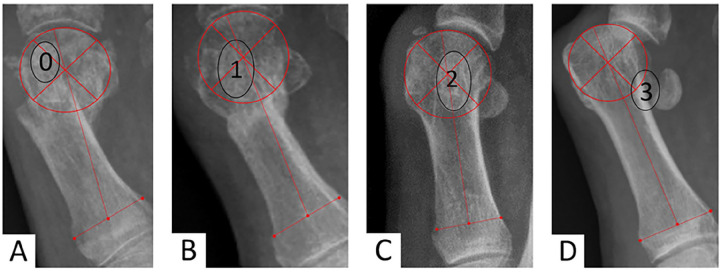
Methods: This retrospective study analyzed 116 feet that underwent percutaneous distal metatarsal transverse osteotomy with lateral soft tissue release and provisional Kirschner wire fixation, with a mean follow-up of 27.1 months, limited to severe cases (hallux valgus angle [HVA] > 40 degrees). Radiologic assessments included preoperative and postoperative measurements of HVA, intermetatarsal angle (IMA), distal metatarsal articular angle, sesamoid position, first metatarsophalangeal (MTPI) joint congruency, metatarsal length, and sagittal position. Clinical evaluations used the AOFAS scale, documenting the recurrence rate, the nature of complications, reoperations, and the association between them. Patient satisfaction was assessed through self-reported evaluations.
Results: Significant improvements were noted for HVA (median correction from 43.1 to 14.6 degrees) and IMA (median correction from 17.2 to 8.5 degrees). The metatarsal was shortened by 5.4 mm. There was a notable reduction in the degree of sesamoid displacement and MTPI congruency. Sagittal position remained unchanged in 85.3%. The median AOFAS score improved from 44.0 to 90.5, well above the clinically significant improvement threshold, and 87.9% of patients were satisfied or very satisfied. We recorded no major complications and minor complications at a rate of 35.3%. Reoperation rate was 14.7%, primarily due to exostoses. Significant associations were found between postoperative sesamoid position and clinical outcome, and between reoperation rate, exostosis, and MTPI congruency, emphasizing the importance of correcting these parameters. Recurrence rate was 6%. Patient satisfaction was associated with reoperation and complications, but not with radiologic parameters.
Conclusion: Percutaneous distal metatarsal osteotomy achieved substantial correction of severe hallux valgus with significant improvements in angular measurements, high patient satisfaction (87.9%), and no major complications. Although the technique shows promise as a less invasive alternative with comparable radiographic outcomes, the 14.7% reoperation rate (primarily for exostoses) and 6% recurrence rate must be considered. Prospective comparative studies are needed to establish its role relative to other surgical approaches for severe deformities.Level of Evidence: Level IV, retrospective case series.


 求助内容:
求助内容: 应助结果提醒方式:


