Sara Oliveira, Pedro Guimarães, Ângelo Roque-Rosado, Elisa Julião Campos, Pedro Serranho, Paulo Matafome, Rui Bernardes, António Francisco Ambrósio
{"title":"基于纹理的OCT分析检测2型糖尿病早期视网膜病变:亚临床糖尿病视网膜病变诊断的潜在方法。","authors":"Sara Oliveira, Pedro Guimarães, Ângelo Roque-Rosado, Elisa Julião Campos, Pedro Serranho, Paulo Matafome, Rui Bernardes, António Francisco Ambrósio","doi":"10.1186/s40662-025-00451-3","DOIUrl":null,"url":null,"abstract":"<p><strong>Background: </strong>Diabetic retinopathy (DR) is often diagnosed many years after diabetes onset, highlighting the need for early diagnosis. The current study aimed to assess whether texture analysis of computed optical coherence tomography (OCT) retinal images can identify (very) early retinal changes. We previously reported retinal texture changes in a type 1 diabetes animal model. This study extends this approach to a type 2 diabetes model exhibiting subtler, more gradually developing retinal alterations to further explore its potential for detecting texture changes when DR-related retinal alterations are minor, strengthening its promising value.</p><p><strong>Methods: </strong>OCT scans and electroretinograms were acquired at baseline and 4, 8, and 12 weeks after initiating the diabetes induction protocol. Automated OCT segmentation, retinal thickness computation, and texture analysis were performed. Blood-retinal barrier permeability, glial reactivity, neuroinflammation, and nitrosative stress were assessed.</p><p><strong>Results: </strong>Retinal texture was affected in the inner plexiform layer and inner/outer photoreceptor segments. At weeks 8 and 12, autocorrelation, cluster prominence, correlation, homogeneity, information measure of correlation II, inverse difference moment normalised, inverse difference normalised, and sum average texture metrics significantly increased/decreased. Importantly, seven of these metrics were also altered in our previous study with type 1 diabetic animals. Type 2 diabetic retinas presented subtle thinning and impaired function, along with a slight reduction in tight junction proteins immunoreactivity, without affecting the blood-retinal barrier.</p><p><strong>Conclusions: </strong>The findings from this study indicate that texture analysis can identify subtle retinal changes during early, clinically silent stages of disease, when biological alterations remain minimal. This highlights its potential utility for the early diagnosis of diabetic retinopathy, though further clinical validation is needed.</p>","PeriodicalId":12194,"journal":{"name":"Eye and Vision","volume":"12 1","pages":"36"},"PeriodicalIF":4.0000,"publicationDate":"2025-09-03","publicationTypes":"Journal Article","fieldsOfStudy":null,"isOpenAccess":false,"openAccessPdf":"https://www.ncbi.nlm.nih.gov/pmc/articles/PMC12406546/pdf/","citationCount":"0","resultStr":"{\"title\":\"Early retinal changes in type 2 diabetes detected by texture-based OCT analysis: potential approach for subclinical diabetic retinopathy diagnosis.\",\"authors\":\"Sara Oliveira, Pedro Guimarães, Ângelo Roque-Rosado, Elisa Julião Campos, Pedro Serranho, Paulo Matafome, Rui Bernardes, António Francisco Ambrósio\",\"doi\":\"10.1186/s40662-025-00451-3\",\"DOIUrl\":null,\"url\":null,\"abstract\":\"<p><strong>Background: </strong>Diabetic retinopathy (DR) is often diagnosed many years after diabetes onset, highlighting the need for early diagnosis. The current study aimed to assess whether texture analysis of computed optical coherence tomography (OCT) retinal images can identify (very) early retinal changes. We previously reported retinal texture changes in a type 1 diabetes animal model. This study extends this approach to a type 2 diabetes model exhibiting subtler, more gradually developing retinal alterations to further explore its potential for detecting texture changes when DR-related retinal alterations are minor, strengthening its promising value.</p><p><strong>Methods: </strong>OCT scans and electroretinograms were acquired at baseline and 4, 8, and 12 weeks after initiating the diabetes induction protocol. Automated OCT segmentation, retinal thickness computation, and texture analysis were performed. Blood-retinal barrier permeability, glial reactivity, neuroinflammation, and nitrosative stress were assessed.</p><p><strong>Results: </strong>Retinal texture was affected in the inner plexiform layer and inner/outer photoreceptor segments. At weeks 8 and 12, autocorrelation, cluster prominence, correlation, homogeneity, information measure of correlation II, inverse difference moment normalised, inverse difference normalised, and sum average texture metrics significantly increased/decreased. Importantly, seven of these metrics were also altered in our previous study with type 1 diabetic animals. Type 2 diabetic retinas presented subtle thinning and impaired function, along with a slight reduction in tight junction proteins immunoreactivity, without affecting the blood-retinal barrier.</p><p><strong>Conclusions: </strong>The findings from this study indicate that texture analysis can identify subtle retinal changes during early, clinically silent stages of disease, when biological alterations remain minimal. This highlights its potential utility for the early diagnosis of diabetic retinopathy, though further clinical validation is needed.</p>\",\"PeriodicalId\":12194,\"journal\":{\"name\":\"Eye and Vision\",\"volume\":\"12 1\",\"pages\":\"36\"},\"PeriodicalIF\":4.0000,\"publicationDate\":\"2025-09-03\",\"publicationTypes\":\"Journal Article\",\"fieldsOfStudy\":null,\"isOpenAccess\":false,\"openAccessPdf\":\"https://www.ncbi.nlm.nih.gov/pmc/articles/PMC12406546/pdf/\",\"citationCount\":\"0\",\"resultStr\":null,\"platform\":\"Semanticscholar\",\"paperid\":null,\"PeriodicalName\":\"Eye and Vision\",\"FirstCategoryId\":\"3\",\"ListUrlMain\":\"https://doi.org/10.1186/s40662-025-00451-3\",\"RegionNum\":1,\"RegionCategory\":\"医学\",\"ArticlePicture\":[],\"TitleCN\":null,\"AbstractTextCN\":null,\"PMCID\":null,\"EPubDate\":\"\",\"PubModel\":\"\",\"JCR\":\"Q1\",\"JCRName\":\"OPHTHALMOLOGY\",\"Score\":null,\"Total\":0}","platform":"Semanticscholar","paperid":null,"PeriodicalName":"Eye and Vision","FirstCategoryId":"3","ListUrlMain":"https://doi.org/10.1186/s40662-025-00451-3","RegionNum":1,"RegionCategory":"医学","ArticlePicture":[],"TitleCN":null,"AbstractTextCN":null,"PMCID":null,"EPubDate":"","PubModel":"","JCR":"Q1","JCRName":"OPHTHALMOLOGY","Score":null,"Total":0}
Early retinal changes in type 2 diabetes detected by texture-based OCT analysis: potential approach for subclinical diabetic retinopathy diagnosis.
Background: Diabetic retinopathy (DR) is often diagnosed many years after diabetes onset, highlighting the need for early diagnosis. The current study aimed to assess whether texture analysis of computed optical coherence tomography (OCT) retinal images can identify (very) early retinal changes. We previously reported retinal texture changes in a type 1 diabetes animal model. This study extends this approach to a type 2 diabetes model exhibiting subtler, more gradually developing retinal alterations to further explore its potential for detecting texture changes when DR-related retinal alterations are minor, strengthening its promising value.
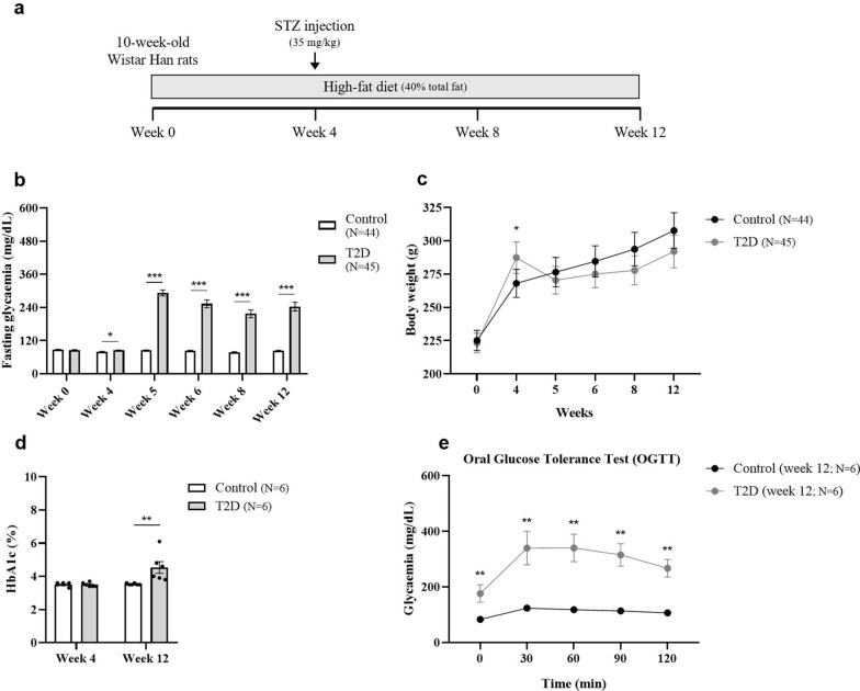
Methods: OCT scans and electroretinograms were acquired at baseline and 4, 8, and 12 weeks after initiating the diabetes induction protocol. Automated OCT segmentation, retinal thickness computation, and texture analysis were performed. Blood-retinal barrier permeability, glial reactivity, neuroinflammation, and nitrosative stress were assessed.
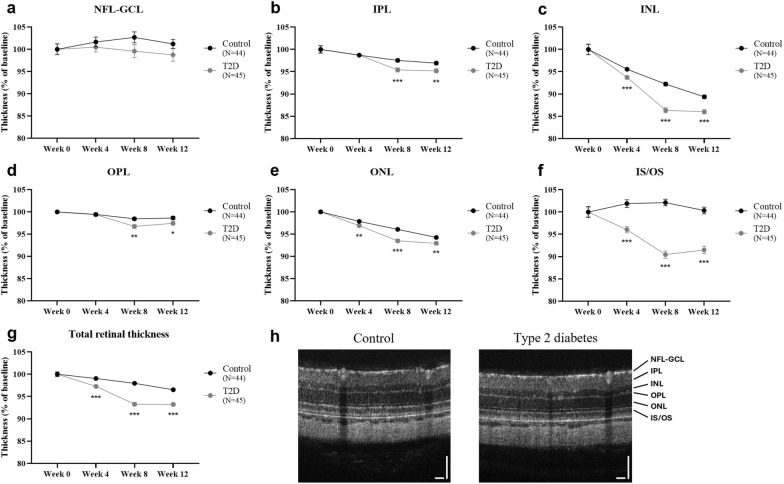
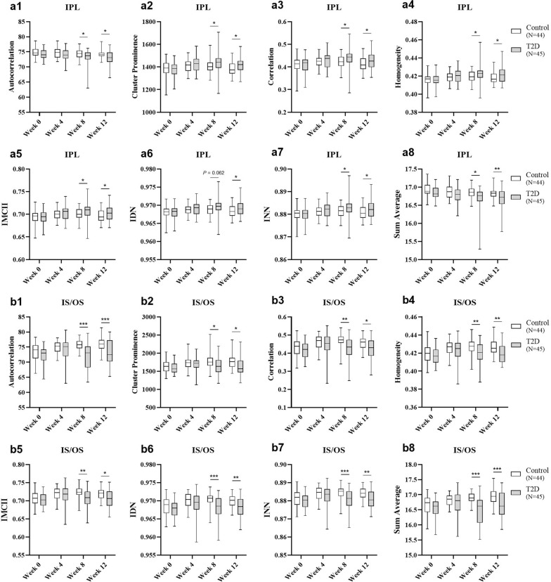
Results: Retinal texture was affected in the inner plexiform layer and inner/outer photoreceptor segments. At weeks 8 and 12, autocorrelation, cluster prominence, correlation, homogeneity, information measure of correlation II, inverse difference moment normalised, inverse difference normalised, and sum average texture metrics significantly increased/decreased. Importantly, seven of these metrics were also altered in our previous study with type 1 diabetic animals. Type 2 diabetic retinas presented subtle thinning and impaired function, along with a slight reduction in tight junction proteins immunoreactivity, without affecting the blood-retinal barrier.
Conclusions: The findings from this study indicate that texture analysis can identify subtle retinal changes during early, clinically silent stages of disease, when biological alterations remain minimal. This highlights its potential utility for the early diagnosis of diabetic retinopathy, though further clinical validation is needed.
期刊介绍:
Eye and Vision is an open access, peer-reviewed journal for ophthalmologists and visual science specialists. It welcomes research articles, reviews, methodologies, commentaries, case reports, perspectives and short reports encompassing all aspects of eye and vision. Topics of interest include but are not limited to: current developments of theoretical, experimental and clinical investigations in ophthalmology, optometry and vision science which focus on novel and high-impact findings on central issues pertaining to biology, pathophysiology and etiology of eye diseases as well as advances in diagnostic techniques, surgical treatment, instrument updates, the latest drug findings, results of clinical trials and research findings. It aims to provide ophthalmologists and visual science specialists with the latest developments in theoretical, experimental and clinical investigations in eye and vision.

 求助内容:
求助内容: 应助结果提醒方式:


