{"title":"基于连接体的脑卒中视觉空间类比推理功能磁共振成像研究。","authors":"Takamichi Tohyama, Masaki Fukunaga, Yohei Otaka","doi":"10.23736/S1973-9087.25.08872-0","DOIUrl":null,"url":null,"abstract":"<p><strong>Background: </strong>Visuospatial function is a core domain of functional cognition in stroke. Post-stroke cognitive impairment disrupts rehabilitation practice, highlighting the importance of characterizing patients with higher-order visuospatial dysfunction to inform personalized rehabilitation strategies. Although neuroimaging offers insights into disease-related mechanisms, its clinical application remains limited.</p><p><strong>Aim: </strong>The aim of this paper was to investigate whether the residual resting-state functional connectivity supports higher-order visuospatial function after stroke and whether changes in connectivity can characterize patients with visuospatial dysfunction.</p><p><strong>Design: </strong>Observational study.</p><p><strong>Setting: </strong>Inpatient rehabilitation ward at Fujita Health University Hospital in Japan.</p><p><strong>Population: </strong>Fifty-eight patients with stroke.</p><p><strong>Methods: </strong>Visuospatial analogical reasoning was assessed using Raven's Colored Progressive Matrices (RCPM). Resting-state functional connectivity was evaluated using functional magnetic resonance imaging (fMRI). Empirical covariance matrices and group-sparse inverse covariance (GSIC) matrices were computed from the fMRI data, with the latter negated to estimate partial correlations representing direct connectivity. Correlations between connectivity measures and RCPM scores were analyzed, alongside data-driven clustering to stratify patients.</p><p><strong>Results: </strong>No significant correlation was found between empirical covariance connectivity and RCPM scores. However, GSIC-based analysis revealed a significant inverse correlation between connectivity of the posteromedial and the left inferior parietal cortex and RCPM scores. Higher parietal connectivity was associated with lower RCPM performance. Patients in the highest connectivity cluster exhibited severe impairments in visuospatial analogical reasoning, particularly in tasks requiring the integration of discrete figures into spatially related wholes. The lesions in these patients were predominantly localized in the left subcortex.</p><p><strong>Conclusions: </strong>Medio-lateral parietal connectivity may underlie visuospatial analogical reasoning after stroke.</p><p><strong>Clinical rehabilitation impact: </strong>Clustering analysis highlighted a distinct pattern of low scores in patients with increased parietal connectivity, suggesting that parietal connectivity changes have the potential for characterizing patients with severe dysfunction.</p>","PeriodicalId":12044,"journal":{"name":"European journal of physical and rehabilitation medicine","volume":"61 3","pages":"462-471"},"PeriodicalIF":3.4000,"publicationDate":"2025-06-01","publicationTypes":"Journal Article","fieldsOfStudy":null,"isOpenAccess":false,"openAccessPdf":"https://www.ncbi.nlm.nih.gov/pmc/articles/PMC12406952/pdf/","citationCount":"0","resultStr":"{\"title\":\"A connectome-based functional magnetic resonance imaging study of visuospatial analogical reasoning in stroke.\",\"authors\":\"Takamichi Tohyama, Masaki Fukunaga, Yohei Otaka\",\"doi\":\"10.23736/S1973-9087.25.08872-0\",\"DOIUrl\":null,\"url\":null,\"abstract\":\"<p><strong>Background: </strong>Visuospatial function is a core domain of functional cognition in stroke. Post-stroke cognitive impairment disrupts rehabilitation practice, highlighting the importance of characterizing patients with higher-order visuospatial dysfunction to inform personalized rehabilitation strategies. Although neuroimaging offers insights into disease-related mechanisms, its clinical application remains limited.</p><p><strong>Aim: </strong>The aim of this paper was to investigate whether the residual resting-state functional connectivity supports higher-order visuospatial function after stroke and whether changes in connectivity can characterize patients with visuospatial dysfunction.</p><p><strong>Design: </strong>Observational study.</p><p><strong>Setting: </strong>Inpatient rehabilitation ward at Fujita Health University Hospital in Japan.</p><p><strong>Population: </strong>Fifty-eight patients with stroke.</p><p><strong>Methods: </strong>Visuospatial analogical reasoning was assessed using Raven's Colored Progressive Matrices (RCPM). Resting-state functional connectivity was evaluated using functional magnetic resonance imaging (fMRI). Empirical covariance matrices and group-sparse inverse covariance (GSIC) matrices were computed from the fMRI data, with the latter negated to estimate partial correlations representing direct connectivity. Correlations between connectivity measures and RCPM scores were analyzed, alongside data-driven clustering to stratify patients.</p><p><strong>Results: </strong>No significant correlation was found between empirical covariance connectivity and RCPM scores. However, GSIC-based analysis revealed a significant inverse correlation between connectivity of the posteromedial and the left inferior parietal cortex and RCPM scores. Higher parietal connectivity was associated with lower RCPM performance. Patients in the highest connectivity cluster exhibited severe impairments in visuospatial analogical reasoning, particularly in tasks requiring the integration of discrete figures into spatially related wholes. The lesions in these patients were predominantly localized in the left subcortex.</p><p><strong>Conclusions: </strong>Medio-lateral parietal connectivity may underlie visuospatial analogical reasoning after stroke.</p><p><strong>Clinical rehabilitation impact: </strong>Clustering analysis highlighted a distinct pattern of low scores in patients with increased parietal connectivity, suggesting that parietal connectivity changes have the potential for characterizing patients with severe dysfunction.</p>\",\"PeriodicalId\":12044,\"journal\":{\"name\":\"European journal of physical and rehabilitation medicine\",\"volume\":\"61 3\",\"pages\":\"462-471\"},\"PeriodicalIF\":3.4000,\"publicationDate\":\"2025-06-01\",\"publicationTypes\":\"Journal Article\",\"fieldsOfStudy\":null,\"isOpenAccess\":false,\"openAccessPdf\":\"https://www.ncbi.nlm.nih.gov/pmc/articles/PMC12406952/pdf/\",\"citationCount\":\"0\",\"resultStr\":null,\"platform\":\"Semanticscholar\",\"paperid\":null,\"PeriodicalName\":\"European journal of physical and rehabilitation medicine\",\"FirstCategoryId\":\"3\",\"ListUrlMain\":\"https://doi.org/10.23736/S1973-9087.25.08872-0\",\"RegionNum\":3,\"RegionCategory\":\"医学\",\"ArticlePicture\":[],\"TitleCN\":null,\"AbstractTextCN\":null,\"PMCID\":null,\"EPubDate\":\"\",\"PubModel\":\"\",\"JCR\":\"Q1\",\"JCRName\":\"REHABILITATION\",\"Score\":null,\"Total\":0}","platform":"Semanticscholar","paperid":null,"PeriodicalName":"European journal of physical and rehabilitation medicine","FirstCategoryId":"3","ListUrlMain":"https://doi.org/10.23736/S1973-9087.25.08872-0","RegionNum":3,"RegionCategory":"医学","ArticlePicture":[],"TitleCN":null,"AbstractTextCN":null,"PMCID":null,"EPubDate":"","PubModel":"","JCR":"Q1","JCRName":"REHABILITATION","Score":null,"Total":0}
A connectome-based functional magnetic resonance imaging study of visuospatial analogical reasoning in stroke.
Background: Visuospatial function is a core domain of functional cognition in stroke. Post-stroke cognitive impairment disrupts rehabilitation practice, highlighting the importance of characterizing patients with higher-order visuospatial dysfunction to inform personalized rehabilitation strategies. Although neuroimaging offers insights into disease-related mechanisms, its clinical application remains limited.
Aim: The aim of this paper was to investigate whether the residual resting-state functional connectivity supports higher-order visuospatial function after stroke and whether changes in connectivity can characterize patients with visuospatial dysfunction.
Design: Observational study.
Setting: Inpatient rehabilitation ward at Fujita Health University Hospital in Japan.
Population: Fifty-eight patients with stroke.
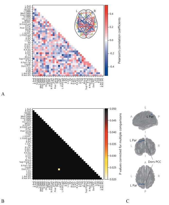
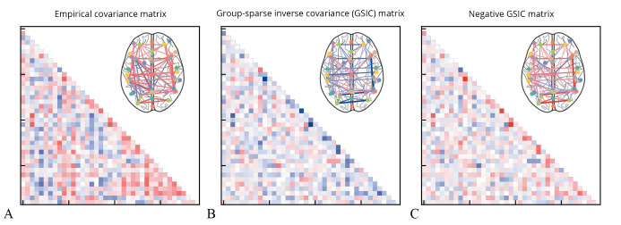
Methods: Visuospatial analogical reasoning was assessed using Raven's Colored Progressive Matrices (RCPM). Resting-state functional connectivity was evaluated using functional magnetic resonance imaging (fMRI). Empirical covariance matrices and group-sparse inverse covariance (GSIC) matrices were computed from the fMRI data, with the latter negated to estimate partial correlations representing direct connectivity. Correlations between connectivity measures and RCPM scores were analyzed, alongside data-driven clustering to stratify patients.
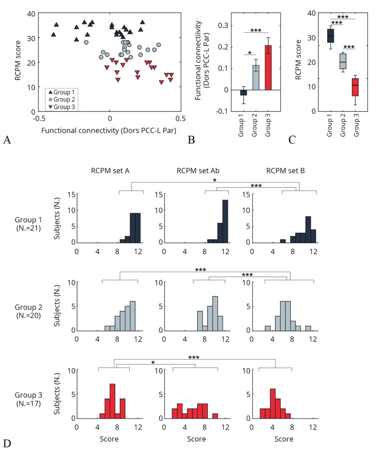
Results: No significant correlation was found between empirical covariance connectivity and RCPM scores. However, GSIC-based analysis revealed a significant inverse correlation between connectivity of the posteromedial and the left inferior parietal cortex and RCPM scores. Higher parietal connectivity was associated with lower RCPM performance. Patients in the highest connectivity cluster exhibited severe impairments in visuospatial analogical reasoning, particularly in tasks requiring the integration of discrete figures into spatially related wholes. The lesions in these patients were predominantly localized in the left subcortex.
Conclusions: Medio-lateral parietal connectivity may underlie visuospatial analogical reasoning after stroke.
Clinical rehabilitation impact: Clustering analysis highlighted a distinct pattern of low scores in patients with increased parietal connectivity, suggesting that parietal connectivity changes have the potential for characterizing patients with severe dysfunction.
 求助内容:
求助内容: 应助结果提醒方式:


