Filippo Migliorini, Nicola Maffulli, Daniel Kämmer, Ulf Krister Hofmann, Jörg Eschweiler, Andreas Bell
{"title":"内侧单室膝关节置换术失败的冠状轴偏差:无菌性松动患者的影像学研究。","authors":"Filippo Migliorini, Nicola Maffulli, Daniel Kämmer, Ulf Krister Hofmann, Jörg Eschweiler, Andreas Bell","doi":"10.1186/s40001-025-03112-2","DOIUrl":null,"url":null,"abstract":"<p><strong>Purpose: </strong>Aseptic loosening remains a leading cause of revision in medial unicompartmental knee arthroplasty (UKA). This imaging study aimed to identify recurrent patterns of coronal alignment deviation in patients undergoing revision to total knee arthroplasty (TKA) to explore whether subtle malalignment may contribute to biomechanical failure.</p><p><strong>Methods: </strong>Imaging of patients who underwent revision surgery of a medial UKA to TKA for aseptic loosening of the tibial or femoral component was retrieved. Lower limb axes were evaluated using anteroposterior plain radiographs of the leg using the software MediCAD Knie 2D (mediCAD Hectec GmbH, Altdorf, Germany). The radiographic axes of revised patients were compared with established reference values, as defined by the MediCAD Knie 2D software and published literature, to identify common alignment patterns potentially associated with aseptic loosening.</p><p><strong>Results: </strong>Data from 62 patients were analysed. Before the revision surgery, the joint line convergence angle (JLCA, P = 0.002) and the anatomical-mechanical angle (AMA, P < 0.0001) were statistically significantly greater than the corresponding reference values. In contrast, the mechanical lateral distal femoral angle (mLDFA, P < 0.0001), the mechanical and anatomical medial proximal tibial angle (mMPTA and aMPTA, P < 0.0001), and the mechanical and anatomical lateral distal tibial angle (mLDTA and aLDTA, P < 0.0001) were significantly lower than reference. No statistically significant difference was found in the mechanical lateral proximal femoral angle (mLPFA, P = 0.9) or in the mechanical axis deviation (MAD, P = 0.5) when compared to normative data.</p><p><strong>Conclusion: </strong>Our cohort of patients revised from medial UKA to TKA for aseptic loosening frequently exhibited consistent deviations in lower limb alignment, particularly increased AMA and JLCA, and reduced mLDFA, mMPTA, and mLDTA. These subtle but recurrent patterns may alter load distribution across the medial compartment, contributing to implant micromotion and loosening. A detailed preoperative axis assessment may help identify patients at a higher biomechanical risk.</p>","PeriodicalId":11949,"journal":{"name":"European Journal of Medical Research","volume":"30 1","pages":"832"},"PeriodicalIF":3.4000,"publicationDate":"2025-09-02","publicationTypes":"Journal Article","fieldsOfStudy":null,"isOpenAccess":false,"openAccessPdf":"https://www.ncbi.nlm.nih.gov/pmc/articles/PMC12403283/pdf/","citationCount":"0","resultStr":"{\"title\":\"Coronal axis deviations in medial unicompartmental knee arthroplasty failures: an imaging study of patients revised for aseptic loosening.\",\"authors\":\"Filippo Migliorini, Nicola Maffulli, Daniel Kämmer, Ulf Krister Hofmann, Jörg Eschweiler, Andreas Bell\",\"doi\":\"10.1186/s40001-025-03112-2\",\"DOIUrl\":null,\"url\":null,\"abstract\":\"<p><strong>Purpose: </strong>Aseptic loosening remains a leading cause of revision in medial unicompartmental knee arthroplasty (UKA). This imaging study aimed to identify recurrent patterns of coronal alignment deviation in patients undergoing revision to total knee arthroplasty (TKA) to explore whether subtle malalignment may contribute to biomechanical failure.</p><p><strong>Methods: </strong>Imaging of patients who underwent revision surgery of a medial UKA to TKA for aseptic loosening of the tibial or femoral component was retrieved. Lower limb axes were evaluated using anteroposterior plain radiographs of the leg using the software MediCAD Knie 2D (mediCAD Hectec GmbH, Altdorf, Germany). The radiographic axes of revised patients were compared with established reference values, as defined by the MediCAD Knie 2D software and published literature, to identify common alignment patterns potentially associated with aseptic loosening.</p><p><strong>Results: </strong>Data from 62 patients were analysed. Before the revision surgery, the joint line convergence angle (JLCA, P = 0.002) and the anatomical-mechanical angle (AMA, P < 0.0001) were statistically significantly greater than the corresponding reference values. In contrast, the mechanical lateral distal femoral angle (mLDFA, P < 0.0001), the mechanical and anatomical medial proximal tibial angle (mMPTA and aMPTA, P < 0.0001), and the mechanical and anatomical lateral distal tibial angle (mLDTA and aLDTA, P < 0.0001) were significantly lower than reference. No statistically significant difference was found in the mechanical lateral proximal femoral angle (mLPFA, P = 0.9) or in the mechanical axis deviation (MAD, P = 0.5) when compared to normative data.</p><p><strong>Conclusion: </strong>Our cohort of patients revised from medial UKA to TKA for aseptic loosening frequently exhibited consistent deviations in lower limb alignment, particularly increased AMA and JLCA, and reduced mLDFA, mMPTA, and mLDTA. These subtle but recurrent patterns may alter load distribution across the medial compartment, contributing to implant micromotion and loosening. A detailed preoperative axis assessment may help identify patients at a higher biomechanical risk.</p>\",\"PeriodicalId\":11949,\"journal\":{\"name\":\"European Journal of Medical Research\",\"volume\":\"30 1\",\"pages\":\"832\"},\"PeriodicalIF\":3.4000,\"publicationDate\":\"2025-09-02\",\"publicationTypes\":\"Journal Article\",\"fieldsOfStudy\":null,\"isOpenAccess\":false,\"openAccessPdf\":\"https://www.ncbi.nlm.nih.gov/pmc/articles/PMC12403283/pdf/\",\"citationCount\":\"0\",\"resultStr\":null,\"platform\":\"Semanticscholar\",\"paperid\":null,\"PeriodicalName\":\"European Journal of Medical Research\",\"FirstCategoryId\":\"3\",\"ListUrlMain\":\"https://doi.org/10.1186/s40001-025-03112-2\",\"RegionNum\":3,\"RegionCategory\":\"医学\",\"ArticlePicture\":[],\"TitleCN\":null,\"AbstractTextCN\":null,\"PMCID\":null,\"EPubDate\":\"\",\"PubModel\":\"\",\"JCR\":\"Q2\",\"JCRName\":\"MEDICINE, RESEARCH & EXPERIMENTAL\",\"Score\":null,\"Total\":0}","platform":"Semanticscholar","paperid":null,"PeriodicalName":"European Journal of Medical Research","FirstCategoryId":"3","ListUrlMain":"https://doi.org/10.1186/s40001-025-03112-2","RegionNum":3,"RegionCategory":"医学","ArticlePicture":[],"TitleCN":null,"AbstractTextCN":null,"PMCID":null,"EPubDate":"","PubModel":"","JCR":"Q2","JCRName":"MEDICINE, RESEARCH & EXPERIMENTAL","Score":null,"Total":0}
Coronal axis deviations in medial unicompartmental knee arthroplasty failures: an imaging study of patients revised for aseptic loosening.
Purpose: Aseptic loosening remains a leading cause of revision in medial unicompartmental knee arthroplasty (UKA). This imaging study aimed to identify recurrent patterns of coronal alignment deviation in patients undergoing revision to total knee arthroplasty (TKA) to explore whether subtle malalignment may contribute to biomechanical failure.
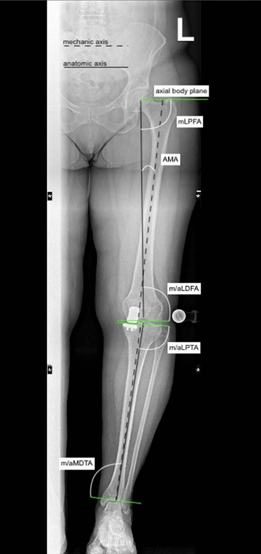
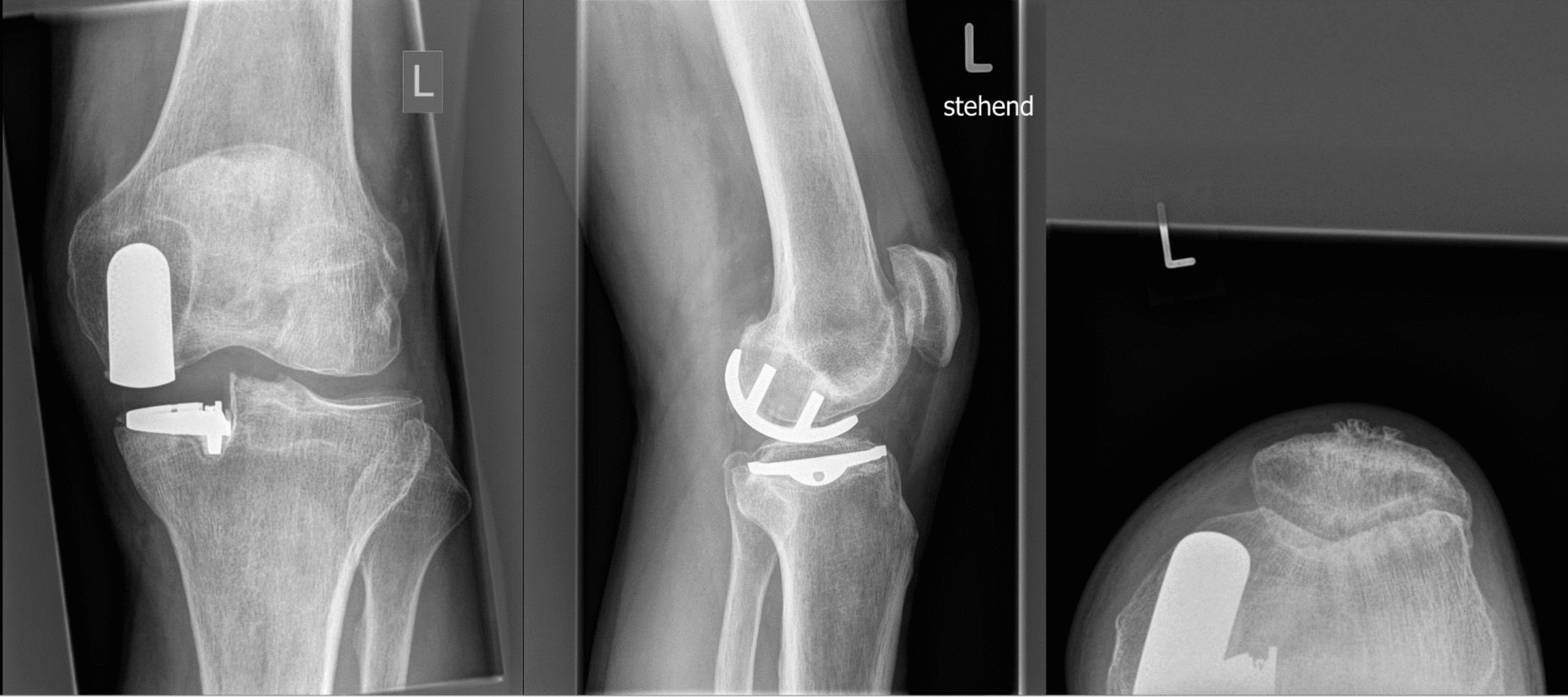
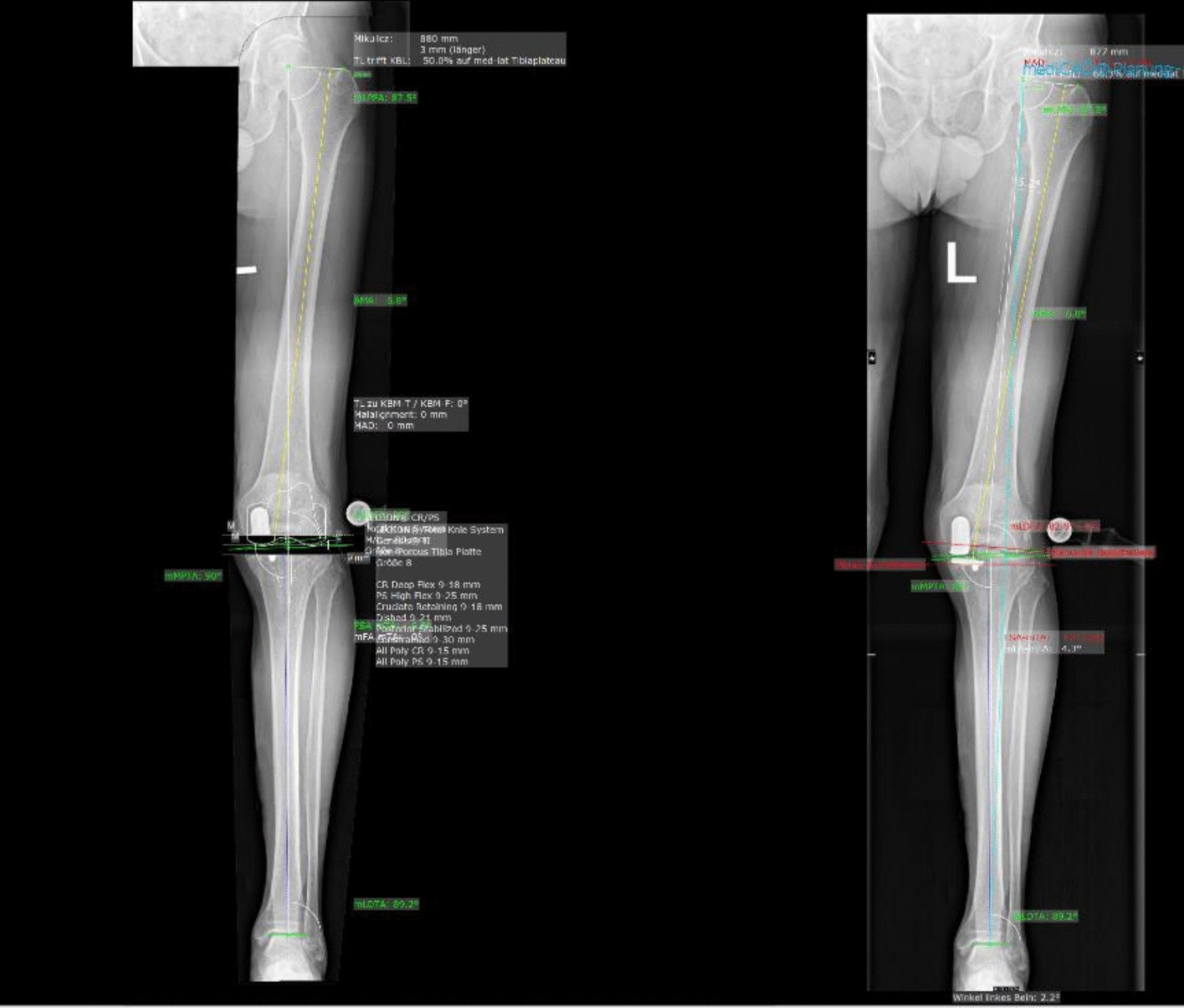
Methods: Imaging of patients who underwent revision surgery of a medial UKA to TKA for aseptic loosening of the tibial or femoral component was retrieved. Lower limb axes were evaluated using anteroposterior plain radiographs of the leg using the software MediCAD Knie 2D (mediCAD Hectec GmbH, Altdorf, Germany). The radiographic axes of revised patients were compared with established reference values, as defined by the MediCAD Knie 2D software and published literature, to identify common alignment patterns potentially associated with aseptic loosening.
Results: Data from 62 patients were analysed. Before the revision surgery, the joint line convergence angle (JLCA, P = 0.002) and the anatomical-mechanical angle (AMA, P < 0.0001) were statistically significantly greater than the corresponding reference values. In contrast, the mechanical lateral distal femoral angle (mLDFA, P < 0.0001), the mechanical and anatomical medial proximal tibial angle (mMPTA and aMPTA, P < 0.0001), and the mechanical and anatomical lateral distal tibial angle (mLDTA and aLDTA, P < 0.0001) were significantly lower than reference. No statistically significant difference was found in the mechanical lateral proximal femoral angle (mLPFA, P = 0.9) or in the mechanical axis deviation (MAD, P = 0.5) when compared to normative data.
Conclusion: Our cohort of patients revised from medial UKA to TKA for aseptic loosening frequently exhibited consistent deviations in lower limb alignment, particularly increased AMA and JLCA, and reduced mLDFA, mMPTA, and mLDTA. These subtle but recurrent patterns may alter load distribution across the medial compartment, contributing to implant micromotion and loosening. A detailed preoperative axis assessment may help identify patients at a higher biomechanical risk.
期刊介绍:
European Journal of Medical Research publishes translational and clinical research of international interest across all medical disciplines, enabling clinicians and other researchers to learn about developments and innovations within these disciplines and across the boundaries between disciplines. The journal publishes high quality research and reviews and aims to ensure that the results of all well-conducted research are published, regardless of their outcome.

 求助内容:
求助内容: 应助结果提醒方式:


