Clara Isabel Perez, Lore Lakunza, Alberto Saenz, Roberto Voces, Ignacio Gallo
{"title":"主动脉弓内动静脉瘘:应用三维模型进行手术计划一例报告。","authors":"Clara Isabel Perez, Lore Lakunza, Alberto Saenz, Roberto Voces, Ignacio Gallo","doi":"10.1093/ehjcr/ytaf362","DOIUrl":null,"url":null,"abstract":"<p><strong>Background: </strong>Traumatic arteriovenous fistulas involving the thoracic aorta are rare. Their closure remains a challenge for the surgeon due to the vascular changes involved. We aim to highlight the benefits of 3D models and virtual three-dimensional reconstruction when used as a complementary tool in surgical planning.</p><p><strong>Case summary: </strong>We present the case of a 38-year-old male patient with a history trauma from a localized bombing, resulting in shrapnel impact to his thorax. He came for surgical treatment due to a diagnosis of arteriovenous fistula at the level of the aortic arch. For surgical planning, two 3D models (with FDM and PolyJet technology) were made to assess the anatomical relationship between the aorta, the left brachiocephalic vein, the fistulous tract and the sternum. The patient was successfully operated on.</p><p><strong>Discussion: </strong>Surgical repair of thoracic fistulas is highly complex, requiring a multi-disciplinary approach and advanced imaging for accurate diagnosis and planning. Procedures are often performed under cardiopulmonary bypass and hypothermia to enhance safety, especially in challenging scenarios. These fistulas frequently cause vascular overload and degenerative venous changes, increasing the risk of haemorrhage and complicating dissection. 3D technology significantly improves surgical planning by enabling accurate fistula localization, guiding sternotomy, simulating procedures, and mapping vascular anomalies. PolyJet models, in particular, provide superior anatomical insight. Thus, 3D modelling serves as a complementary tool that provides life-size, detailed anatomical information of the patient, facilitating better pre-operative surgical planning, and carrying the potential to reduce operative risks.</p>","PeriodicalId":11910,"journal":{"name":"European Heart Journal: Case Reports","volume":"9 8","pages":"ytaf362"},"PeriodicalIF":0.8000,"publicationDate":"2025-07-30","publicationTypes":"Journal Article","fieldsOfStudy":null,"isOpenAccess":false,"openAccessPdf":"https://www.ncbi.nlm.nih.gov/pmc/articles/PMC12368333/pdf/","citationCount":"0","resultStr":"{\"title\":\"Arteriovenous fistula in the aortic arch: a case report involving 3D models for surgical planning.\",\"authors\":\"Clara Isabel Perez, Lore Lakunza, Alberto Saenz, Roberto Voces, Ignacio Gallo\",\"doi\":\"10.1093/ehjcr/ytaf362\",\"DOIUrl\":null,\"url\":null,\"abstract\":\"<p><strong>Background: </strong>Traumatic arteriovenous fistulas involving the thoracic aorta are rare. Their closure remains a challenge for the surgeon due to the vascular changes involved. We aim to highlight the benefits of 3D models and virtual three-dimensional reconstruction when used as a complementary tool in surgical planning.</p><p><strong>Case summary: </strong>We present the case of a 38-year-old male patient with a history trauma from a localized bombing, resulting in shrapnel impact to his thorax. He came for surgical treatment due to a diagnosis of arteriovenous fistula at the level of the aortic arch. For surgical planning, two 3D models (with FDM and PolyJet technology) were made to assess the anatomical relationship between the aorta, the left brachiocephalic vein, the fistulous tract and the sternum. The patient was successfully operated on.</p><p><strong>Discussion: </strong>Surgical repair of thoracic fistulas is highly complex, requiring a multi-disciplinary approach and advanced imaging for accurate diagnosis and planning. Procedures are often performed under cardiopulmonary bypass and hypothermia to enhance safety, especially in challenging scenarios. These fistulas frequently cause vascular overload and degenerative venous changes, increasing the risk of haemorrhage and complicating dissection. 3D technology significantly improves surgical planning by enabling accurate fistula localization, guiding sternotomy, simulating procedures, and mapping vascular anomalies. PolyJet models, in particular, provide superior anatomical insight. Thus, 3D modelling serves as a complementary tool that provides life-size, detailed anatomical information of the patient, facilitating better pre-operative surgical planning, and carrying the potential to reduce operative risks.</p>\",\"PeriodicalId\":11910,\"journal\":{\"name\":\"European Heart Journal: Case Reports\",\"volume\":\"9 8\",\"pages\":\"ytaf362\"},\"PeriodicalIF\":0.8000,\"publicationDate\":\"2025-07-30\",\"publicationTypes\":\"Journal Article\",\"fieldsOfStudy\":null,\"isOpenAccess\":false,\"openAccessPdf\":\"https://www.ncbi.nlm.nih.gov/pmc/articles/PMC12368333/pdf/\",\"citationCount\":\"0\",\"resultStr\":null,\"platform\":\"Semanticscholar\",\"paperid\":null,\"PeriodicalName\":\"European Heart Journal: Case Reports\",\"FirstCategoryId\":\"1085\",\"ListUrlMain\":\"https://doi.org/10.1093/ehjcr/ytaf362\",\"RegionNum\":0,\"RegionCategory\":null,\"ArticlePicture\":[],\"TitleCN\":null,\"AbstractTextCN\":null,\"PMCID\":null,\"EPubDate\":\"2025/8/1 0:00:00\",\"PubModel\":\"eCollection\",\"JCR\":\"Q4\",\"JCRName\":\"CARDIAC & CARDIOVASCULAR SYSTEMS\",\"Score\":null,\"Total\":0}","platform":"Semanticscholar","paperid":null,"PeriodicalName":"European Heart Journal: Case Reports","FirstCategoryId":"1085","ListUrlMain":"https://doi.org/10.1093/ehjcr/ytaf362","RegionNum":0,"RegionCategory":null,"ArticlePicture":[],"TitleCN":null,"AbstractTextCN":null,"PMCID":null,"EPubDate":"2025/8/1 0:00:00","PubModel":"eCollection","JCR":"Q4","JCRName":"CARDIAC & CARDIOVASCULAR SYSTEMS","Score":null,"Total":0}
Arteriovenous fistula in the aortic arch: a case report involving 3D models for surgical planning.
Background: Traumatic arteriovenous fistulas involving the thoracic aorta are rare. Their closure remains a challenge for the surgeon due to the vascular changes involved. We aim to highlight the benefits of 3D models and virtual three-dimensional reconstruction when used as a complementary tool in surgical planning.
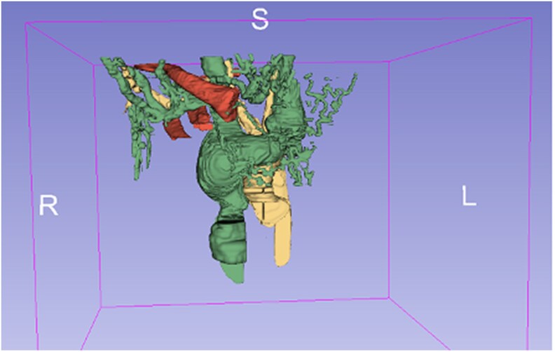
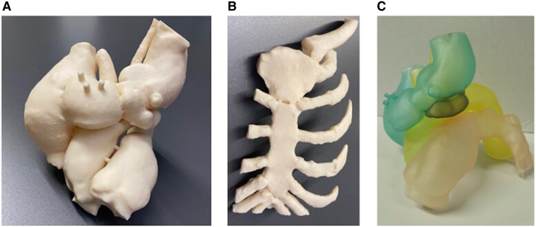
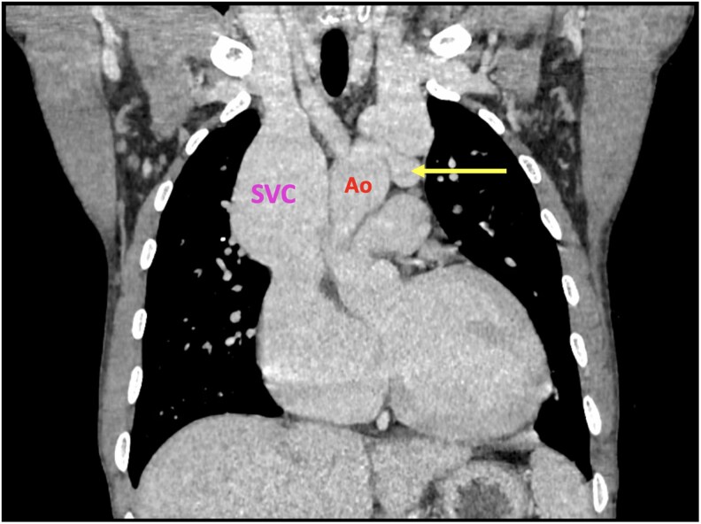
Case summary: We present the case of a 38-year-old male patient with a history trauma from a localized bombing, resulting in shrapnel impact to his thorax. He came for surgical treatment due to a diagnosis of arteriovenous fistula at the level of the aortic arch. For surgical planning, two 3D models (with FDM and PolyJet technology) were made to assess the anatomical relationship between the aorta, the left brachiocephalic vein, the fistulous tract and the sternum. The patient was successfully operated on.
Discussion: Surgical repair of thoracic fistulas is highly complex, requiring a multi-disciplinary approach and advanced imaging for accurate diagnosis and planning. Procedures are often performed under cardiopulmonary bypass and hypothermia to enhance safety, especially in challenging scenarios. These fistulas frequently cause vascular overload and degenerative venous changes, increasing the risk of haemorrhage and complicating dissection. 3D technology significantly improves surgical planning by enabling accurate fistula localization, guiding sternotomy, simulating procedures, and mapping vascular anomalies. PolyJet models, in particular, provide superior anatomical insight. Thus, 3D modelling serves as a complementary tool that provides life-size, detailed anatomical information of the patient, facilitating better pre-operative surgical planning, and carrying the potential to reduce operative risks.

 求助内容:
求助内容: 应助结果提醒方式:


