Pierre Lafeuille, Renato Medas, Benjamin Hamel, Romain Legros, Sarah Leblanc, Maximilien Barret, Vincent Lepilliez, Juliette Leroux, Thimothee Wallenhorst, Dann Joseph Ouizeman, Clement Fortier Beaulieu, Hugo Uchima, Elena De Cristofaro, Yann Le Baleur, Antoine Debourdeau, Fabien Subtil, Tanguy Fenouil, Alexandru Lupu, Florian Rostain, Jérôme Rivory, Jeremie Jacques, João Santos-Antunes, Mathieu Pioche
{"title":"结肠粘膜下病变的组织学显示,高比例的良性病变不需要R0整体内镜切除。","authors":"Pierre Lafeuille, Renato Medas, Benjamin Hamel, Romain Legros, Sarah Leblanc, Maximilien Barret, Vincent Lepilliez, Juliette Leroux, Thimothee Wallenhorst, Dann Joseph Ouizeman, Clement Fortier Beaulieu, Hugo Uchima, Elena De Cristofaro, Yann Le Baleur, Antoine Debourdeau, Fabien Subtil, Tanguy Fenouil, Alexandru Lupu, Florian Rostain, Jérôme Rivory, Jeremie Jacques, João Santos-Antunes, Mathieu Pioche","doi":"10.1055/a-2641-5256","DOIUrl":null,"url":null,"abstract":"<p><strong>Background and study aims: </strong>Submucosal lesions in the colon are much rarer than those in the rectum. Their nature is poorly understood, as is the best technique for their excision. Based on that of rectal lesions, it most often aims for R0 en bloc resection, but without formal proof of efficacy. The aim of this study was to evaluate histology of these lesions and determine whether submucosal lesions of the colon always require R0 en bloc endoscopic resection.</p><p><strong>Patients and methods: </strong>We conducted a retrospective international study of all colonic submucosal lesions with confirmed histology by resection or biopsy. We assessed the proportion of lesions correctly managed by endoscopy, so that the proposed resection technique offered a level of tumor resection quality appropriate to the definitive histology of the lesion.</p><p><strong>Results: </strong>One hundred patients with 105 colonic submucosal lesions from 13 European centers were included. Histology revealed 91.4% (96/105) non-malignant lesions and 8.6% (9/105) malignant lesions. Endoscopic techniques used were curative in 41.7% (5/12) of cases requiring resection, non-curative in 58.3% (7/12), and endoscopic resection was not necessary in 88.7% (93/105). There was no delayed surgery for adverse events.</p><p><strong>Conclusions: </strong>Most colonic submucosal lesions are non-malignant and do not warrant advanced endoscopic resection. A new therapeutic approach could be first-line use of a low-risk, low-cost histological diagnostic technique followed in a second phase by a more advanced technique in the event of a malignant histological result. Further studies are needed to evaluate this step-up strategy.</p>","PeriodicalId":11671,"journal":{"name":"Endoscopy International Open","volume":"13 ","pages":"a26415256"},"PeriodicalIF":2.3000,"publicationDate":"2025-07-24","publicationTypes":"Journal Article","fieldsOfStudy":null,"isOpenAccess":false,"openAccessPdf":"https://www.ncbi.nlm.nih.gov/pmc/articles/PMC12372418/pdf/","citationCount":"0","resultStr":"{\"title\":\"Histology of colonic submucosal lesions reveals a high proportion of benign lesions that do not require R0 en bloc endoscopic resection.\",\"authors\":\"Pierre Lafeuille, Renato Medas, Benjamin Hamel, Romain Legros, Sarah Leblanc, Maximilien Barret, Vincent Lepilliez, Juliette Leroux, Thimothee Wallenhorst, Dann Joseph Ouizeman, Clement Fortier Beaulieu, Hugo Uchima, Elena De Cristofaro, Yann Le Baleur, Antoine Debourdeau, Fabien Subtil, Tanguy Fenouil, Alexandru Lupu, Florian Rostain, Jérôme Rivory, Jeremie Jacques, João Santos-Antunes, Mathieu Pioche\",\"doi\":\"10.1055/a-2641-5256\",\"DOIUrl\":null,\"url\":null,\"abstract\":\"<p><strong>Background and study aims: </strong>Submucosal lesions in the colon are much rarer than those in the rectum. Their nature is poorly understood, as is the best technique for their excision. Based on that of rectal lesions, it most often aims for R0 en bloc resection, but without formal proof of efficacy. The aim of this study was to evaluate histology of these lesions and determine whether submucosal lesions of the colon always require R0 en bloc endoscopic resection.</p><p><strong>Patients and methods: </strong>We conducted a retrospective international study of all colonic submucosal lesions with confirmed histology by resection or biopsy. We assessed the proportion of lesions correctly managed by endoscopy, so that the proposed resection technique offered a level of tumor resection quality appropriate to the definitive histology of the lesion.</p><p><strong>Results: </strong>One hundred patients with 105 colonic submucosal lesions from 13 European centers were included. Histology revealed 91.4% (96/105) non-malignant lesions and 8.6% (9/105) malignant lesions. Endoscopic techniques used were curative in 41.7% (5/12) of cases requiring resection, non-curative in 58.3% (7/12), and endoscopic resection was not necessary in 88.7% (93/105). There was no delayed surgery for adverse events.</p><p><strong>Conclusions: </strong>Most colonic submucosal lesions are non-malignant and do not warrant advanced endoscopic resection. A new therapeutic approach could be first-line use of a low-risk, low-cost histological diagnostic technique followed in a second phase by a more advanced technique in the event of a malignant histological result. Further studies are needed to evaluate this step-up strategy.</p>\",\"PeriodicalId\":11671,\"journal\":{\"name\":\"Endoscopy International Open\",\"volume\":\"13 \",\"pages\":\"a26415256\"},\"PeriodicalIF\":2.3000,\"publicationDate\":\"2025-07-24\",\"publicationTypes\":\"Journal Article\",\"fieldsOfStudy\":null,\"isOpenAccess\":false,\"openAccessPdf\":\"https://www.ncbi.nlm.nih.gov/pmc/articles/PMC12372418/pdf/\",\"citationCount\":\"0\",\"resultStr\":null,\"platform\":\"Semanticscholar\",\"paperid\":null,\"PeriodicalName\":\"Endoscopy International Open\",\"FirstCategoryId\":\"1085\",\"ListUrlMain\":\"https://doi.org/10.1055/a-2641-5256\",\"RegionNum\":0,\"RegionCategory\":null,\"ArticlePicture\":[],\"TitleCN\":null,\"AbstractTextCN\":null,\"PMCID\":null,\"EPubDate\":\"2025/1/1 0:00:00\",\"PubModel\":\"eCollection\",\"JCR\":\"Q3\",\"JCRName\":\"GASTROENTEROLOGY & HEPATOLOGY\",\"Score\":null,\"Total\":0}","platform":"Semanticscholar","paperid":null,"PeriodicalName":"Endoscopy International Open","FirstCategoryId":"1085","ListUrlMain":"https://doi.org/10.1055/a-2641-5256","RegionNum":0,"RegionCategory":null,"ArticlePicture":[],"TitleCN":null,"AbstractTextCN":null,"PMCID":null,"EPubDate":"2025/1/1 0:00:00","PubModel":"eCollection","JCR":"Q3","JCRName":"GASTROENTEROLOGY & HEPATOLOGY","Score":null,"Total":0}
Histology of colonic submucosal lesions reveals a high proportion of benign lesions that do not require R0 en bloc endoscopic resection.
Background and study aims: Submucosal lesions in the colon are much rarer than those in the rectum. Their nature is poorly understood, as is the best technique for their excision. Based on that of rectal lesions, it most often aims for R0 en bloc resection, but without formal proof of efficacy. The aim of this study was to evaluate histology of these lesions and determine whether submucosal lesions of the colon always require R0 en bloc endoscopic resection.
Patients and methods: We conducted a retrospective international study of all colonic submucosal lesions with confirmed histology by resection or biopsy. We assessed the proportion of lesions correctly managed by endoscopy, so that the proposed resection technique offered a level of tumor resection quality appropriate to the definitive histology of the lesion.
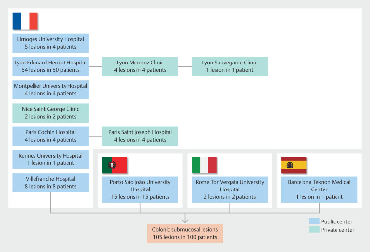
Results: One hundred patients with 105 colonic submucosal lesions from 13 European centers were included. Histology revealed 91.4% (96/105) non-malignant lesions and 8.6% (9/105) malignant lesions. Endoscopic techniques used were curative in 41.7% (5/12) of cases requiring resection, non-curative in 58.3% (7/12), and endoscopic resection was not necessary in 88.7% (93/105). There was no delayed surgery for adverse events.
Conclusions: Most colonic submucosal lesions are non-malignant and do not warrant advanced endoscopic resection. A new therapeutic approach could be first-line use of a low-risk, low-cost histological diagnostic technique followed in a second phase by a more advanced technique in the event of a malignant histological result. Further studies are needed to evaluate this step-up strategy.

 求助内容:
求助内容: 应助结果提醒方式:


