Samir Nammour, Marwan El Mobadder, Aldo Brugnera, Praveen Arany, Mireille El Feghali, Paul Nahas, Alain Vanheusden
{"title":"445nm二极管激光增强牙釉质抗酸性的显微和晶体学分析:离体研究。","authors":"Samir Nammour, Marwan El Mobadder, Aldo Brugnera, Praveen Arany, Mireille El Feghali, Paul Nahas, Alain Vanheusden","doi":"10.3390/dj13080376","DOIUrl":null,"url":null,"abstract":"<p><p><b>Background/Objectives</b>: This study aimed to evaluate the efficacy of a 445 nm diode laser in enhancing enamel resistance to acid-induced demineralization and to investigate the associated compositional and structural modifications using scanning electron microscopy (SEM), electron spectroscopy for chemical analysis (ESCA), and X-ray diffraction (XRD) crystallographic analysis. <b>Methods</b>: A total of 126 extracted human teeth were used. A total of 135 (<i>n</i> = 135) enamel discs (4 × 4 mm) from 90 teeth were assigned to either a laser-irradiated group or an untreated control group for SEM, ESCA, and XRD analyses. Additionally, 24 mono-rooted teeth were used to measure pulp temperature changes during laser application. Laser irradiation was performed using a 445 nm diode laser with a pulse width of 200 ms, a repetition rate of 1 Hz, power of 1.25 W, an energy density of 800 J/cm<sup>2</sup>, a power density of 3980 W/cm<sup>2</sup>, and a 200 µm activated fiber. Following acid etching, SEM was conducted to assess microstructural and ionic alterations. The ESCA was used to evaluate the Ca/P ratio, and XRD analyses were performed on enamel powders to determine changes in phase composition and crystal lattice parameters. <b>Results</b>: The laser protocol demonstrated thermal safety, with minimal pulp chamber temperature elevation (0.05667 ± 0.04131 °C). SEM showed that laser-treated enamel had a smoother surface morphology and reduced acid-induced erosion compared with controls. Results of the ESCA revealed no significant difference in the Ca/P ratio between groups. XRD confirmed the presence of hydroxyapatite structure in laser-treated enamel and detected an additional diffraction peak corresponding to a pyrophosphate phase, potentially enhancing acid resistance. Results of the spectral analysis showed the absence of α-TCP and β-TCP phases and a reduction in the carbonate content in the laser group. Furthermore, a significant decrease in the a-axis lattice parameter suggested lattice compaction in laser-treated enamel. <b>Conclusions</b>: Irradiation with a 445 nm diode laser effectively enhances enamel resistance to acid demineralization. This improvement may be attributed to chemical modifications, particularly pyrophosphate phase formation, and structural changes including prism-less enamel formation, surface fusion, and decreased permeability. These findings provide novel insights into the mechanisms of laser-induced enhancement of acid resistance in enamel.</p>","PeriodicalId":11269,"journal":{"name":"Dentistry Journal","volume":"13 8","pages":""},"PeriodicalIF":3.1000,"publicationDate":"2025-08-19","publicationTypes":"Journal Article","fieldsOfStudy":null,"isOpenAccess":false,"openAccessPdf":"https://www.ncbi.nlm.nih.gov/pmc/articles/PMC12385602/pdf/","citationCount":"0","resultStr":"{\"title\":\"Microscopic and Crystallographic Analysis of Increased Acid Resistance of Melted Dental Enamel Using 445 nm Diode Laser: An Ex-Vivo Study.\",\"authors\":\"Samir Nammour, Marwan El Mobadder, Aldo Brugnera, Praveen Arany, Mireille El Feghali, Paul Nahas, Alain Vanheusden\",\"doi\":\"10.3390/dj13080376\",\"DOIUrl\":null,\"url\":null,\"abstract\":\"<p><p><b>Background/Objectives</b>: This study aimed to evaluate the efficacy of a 445 nm diode laser in enhancing enamel resistance to acid-induced demineralization and to investigate the associated compositional and structural modifications using scanning electron microscopy (SEM), electron spectroscopy for chemical analysis (ESCA), and X-ray diffraction (XRD) crystallographic analysis. <b>Methods</b>: A total of 126 extracted human teeth were used. A total of 135 (<i>n</i> = 135) enamel discs (4 × 4 mm) from 90 teeth were assigned to either a laser-irradiated group or an untreated control group for SEM, ESCA, and XRD analyses. Additionally, 24 mono-rooted teeth were used to measure pulp temperature changes during laser application. Laser irradiation was performed using a 445 nm diode laser with a pulse width of 200 ms, a repetition rate of 1 Hz, power of 1.25 W, an energy density of 800 J/cm<sup>2</sup>, a power density of 3980 W/cm<sup>2</sup>, and a 200 µm activated fiber. Following acid etching, SEM was conducted to assess microstructural and ionic alterations. The ESCA was used to evaluate the Ca/P ratio, and XRD analyses were performed on enamel powders to determine changes in phase composition and crystal lattice parameters. <b>Results</b>: The laser protocol demonstrated thermal safety, with minimal pulp chamber temperature elevation (0.05667 ± 0.04131 °C). SEM showed that laser-treated enamel had a smoother surface morphology and reduced acid-induced erosion compared with controls. Results of the ESCA revealed no significant difference in the Ca/P ratio between groups. XRD confirmed the presence of hydroxyapatite structure in laser-treated enamel and detected an additional diffraction peak corresponding to a pyrophosphate phase, potentially enhancing acid resistance. Results of the spectral analysis showed the absence of α-TCP and β-TCP phases and a reduction in the carbonate content in the laser group. Furthermore, a significant decrease in the a-axis lattice parameter suggested lattice compaction in laser-treated enamel. <b>Conclusions</b>: Irradiation with a 445 nm diode laser effectively enhances enamel resistance to acid demineralization. This improvement may be attributed to chemical modifications, particularly pyrophosphate phase formation, and structural changes including prism-less enamel formation, surface fusion, and decreased permeability. These findings provide novel insights into the mechanisms of laser-induced enhancement of acid resistance in enamel.</p>\",\"PeriodicalId\":11269,\"journal\":{\"name\":\"Dentistry Journal\",\"volume\":\"13 8\",\"pages\":\"\"},\"PeriodicalIF\":3.1000,\"publicationDate\":\"2025-08-19\",\"publicationTypes\":\"Journal Article\",\"fieldsOfStudy\":null,\"isOpenAccess\":false,\"openAccessPdf\":\"https://www.ncbi.nlm.nih.gov/pmc/articles/PMC12385602/pdf/\",\"citationCount\":\"0\",\"resultStr\":null,\"platform\":\"Semanticscholar\",\"paperid\":null,\"PeriodicalName\":\"Dentistry Journal\",\"FirstCategoryId\":\"1085\",\"ListUrlMain\":\"https://doi.org/10.3390/dj13080376\",\"RegionNum\":0,\"RegionCategory\":null,\"ArticlePicture\":[],\"TitleCN\":null,\"AbstractTextCN\":null,\"PMCID\":null,\"EPubDate\":\"\",\"PubModel\":\"\",\"JCR\":\"Q2\",\"JCRName\":\"DENTISTRY, ORAL SURGERY & MEDICINE\",\"Score\":null,\"Total\":0}","platform":"Semanticscholar","paperid":null,"PeriodicalName":"Dentistry Journal","FirstCategoryId":"1085","ListUrlMain":"https://doi.org/10.3390/dj13080376","RegionNum":0,"RegionCategory":null,"ArticlePicture":[],"TitleCN":null,"AbstractTextCN":null,"PMCID":null,"EPubDate":"","PubModel":"","JCR":"Q2","JCRName":"DENTISTRY, ORAL SURGERY & MEDICINE","Score":null,"Total":0}
Microscopic and Crystallographic Analysis of Increased Acid Resistance of Melted Dental Enamel Using 445 nm Diode Laser: An Ex-Vivo Study.
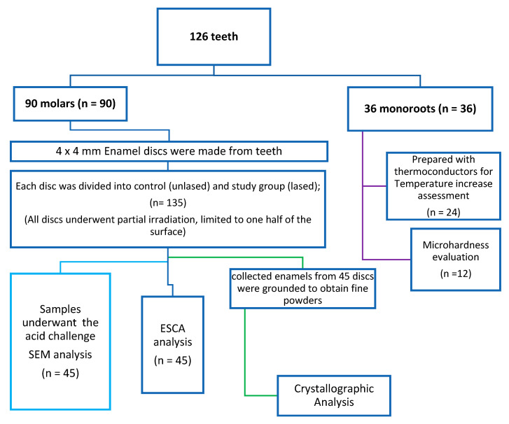
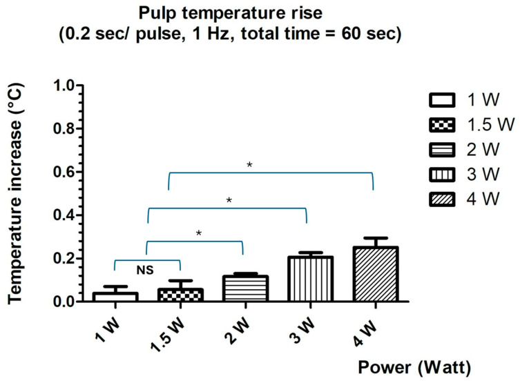
Background/Objectives: This study aimed to evaluate the efficacy of a 445 nm diode laser in enhancing enamel resistance to acid-induced demineralization and to investigate the associated compositional and structural modifications using scanning electron microscopy (SEM), electron spectroscopy for chemical analysis (ESCA), and X-ray diffraction (XRD) crystallographic analysis. Methods: A total of 126 extracted human teeth were used. A total of 135 (n = 135) enamel discs (4 × 4 mm) from 90 teeth were assigned to either a laser-irradiated group or an untreated control group for SEM, ESCA, and XRD analyses. Additionally, 24 mono-rooted teeth were used to measure pulp temperature changes during laser application. Laser irradiation was performed using a 445 nm diode laser with a pulse width of 200 ms, a repetition rate of 1 Hz, power of 1.25 W, an energy density of 800 J/cm2, a power density of 3980 W/cm2, and a 200 µm activated fiber. Following acid etching, SEM was conducted to assess microstructural and ionic alterations. The ESCA was used to evaluate the Ca/P ratio, and XRD analyses were performed on enamel powders to determine changes in phase composition and crystal lattice parameters. Results: The laser protocol demonstrated thermal safety, with minimal pulp chamber temperature elevation (0.05667 ± 0.04131 °C). SEM showed that laser-treated enamel had a smoother surface morphology and reduced acid-induced erosion compared with controls. Results of the ESCA revealed no significant difference in the Ca/P ratio between groups. XRD confirmed the presence of hydroxyapatite structure in laser-treated enamel and detected an additional diffraction peak corresponding to a pyrophosphate phase, potentially enhancing acid resistance. Results of the spectral analysis showed the absence of α-TCP and β-TCP phases and a reduction in the carbonate content in the laser group. Furthermore, a significant decrease in the a-axis lattice parameter suggested lattice compaction in laser-treated enamel. Conclusions: Irradiation with a 445 nm diode laser effectively enhances enamel resistance to acid demineralization. This improvement may be attributed to chemical modifications, particularly pyrophosphate phase formation, and structural changes including prism-less enamel formation, surface fusion, and decreased permeability. These findings provide novel insights into the mechanisms of laser-induced enhancement of acid resistance in enamel.

 求助内容:
求助内容: 应助结果提醒方式:


