{"title":"
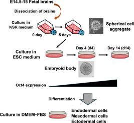
重建小鼠胎儿脑细胞三维结构诱导的多谱系电位。","authors":"Ken Ohnishi , Jun-ichi Sagara , Tomoya Fujita","doi":"10.1016/j.ydbio.2025.08.015","DOIUrl":null,"url":null,"abstract":"<div><div>We examined whether the multilineage potential of mouse fetal brain cells is induced by the three-dimensional (3D) structural reconstruction. Dissociated single cells from the cerebral hemispheres of mouse fetuses at embryonic day 14.5–15 (E14.5–15) rapidly aggregated into cell clusters, forming spherical cell aggregates (SCAs). Immunofluorescence staining revealed that SCAs were positive for Map2 but negative for Nestin, GFAP, and markers of undifferentiated cells such as Oct4, SSEA-1, ALDH2, and CD133. During culture of SCAs in embryonic stem cell medium, SCAs subsequently developed into embryoid bodies (EBs), which began to exhibit positive immunofluorescence staining for Oct4. Reverse transcription–polymerase chain reaction (RT-PCR) and Western blotting analyses further confirmed the expression of Oct4 and Nanog at the gene and protein levels. Differentiated cells of EBs induced by culture in various types of media showed positive immunofluorescence staining for lineage-specific markers, including AFP, α-SMA, GFAP, Map2 and C-peptide and expressed the corresponding genes and proteins. These results suggest that mouse fetal brain cells can regain multilineage potential through the reconstruction of 3D structures without genetic modification. Our findings indicate that 3D culture systems may support non-genetic reprogramming strategies in regenerative medicine by activating intrinsic plasticity of cells.</div></div>","PeriodicalId":11070,"journal":{"name":"Developmental biology","volume":"527 ","pages":"Pages 348-359"},"PeriodicalIF":2.1000,"publicationDate":"2025-08-19","publicationTypes":"Journal Article","fieldsOfStudy":null,"isOpenAccess":false,"openAccessPdf":"","citationCount":"0","resultStr":"{\"title\":\"Multilineage potential induced by reconstruction of three-dimensional structures in mouse fetal brain cells\",\"authors\":\"Ken Ohnishi , Jun-ichi Sagara , Tomoya Fujita\",\"doi\":\"10.1016/j.ydbio.2025.08.015\",\"DOIUrl\":null,\"url\":null,\"abstract\":\"<div><div>We examined whether the multilineage potential of mouse fetal brain cells is induced by the three-dimensional (3D) structural reconstruction. Dissociated single cells from the cerebral hemispheres of mouse fetuses at embryonic day 14.5–15 (E14.5–15) rapidly aggregated into cell clusters, forming spherical cell aggregates (SCAs). Immunofluorescence staining revealed that SCAs were positive for Map2 but negative for Nestin, GFAP, and markers of undifferentiated cells such as Oct4, SSEA-1, ALDH2, and CD133. During culture of SCAs in embryonic stem cell medium, SCAs subsequently developed into embryoid bodies (EBs), which began to exhibit positive immunofluorescence staining for Oct4. Reverse transcription–polymerase chain reaction (RT-PCR) and Western blotting analyses further confirmed the expression of Oct4 and Nanog at the gene and protein levels. Differentiated cells of EBs induced by culture in various types of media showed positive immunofluorescence staining for lineage-specific markers, including AFP, α-SMA, GFAP, Map2 and C-peptide and expressed the corresponding genes and proteins. These results suggest that mouse fetal brain cells can regain multilineage potential through the reconstruction of 3D structures without genetic modification. Our findings indicate that 3D culture systems may support non-genetic reprogramming strategies in regenerative medicine by activating intrinsic plasticity of cells.</div></div>\",\"PeriodicalId\":11070,\"journal\":{\"name\":\"Developmental biology\",\"volume\":\"527 \",\"pages\":\"Pages 348-359\"},\"PeriodicalIF\":2.1000,\"publicationDate\":\"2025-08-19\",\"publicationTypes\":\"Journal Article\",\"fieldsOfStudy\":null,\"isOpenAccess\":false,\"openAccessPdf\":\"\",\"citationCount\":\"0\",\"resultStr\":null,\"platform\":\"Semanticscholar\",\"paperid\":null,\"PeriodicalName\":\"Developmental biology\",\"FirstCategoryId\":\"99\",\"ListUrlMain\":\"https://www.sciencedirect.com/science/article/pii/S0012160625002325\",\"RegionNum\":3,\"RegionCategory\":\"生物学\",\"ArticlePicture\":[],\"TitleCN\":null,\"AbstractTextCN\":null,\"PMCID\":null,\"EPubDate\":\"\",\"PubModel\":\"\",\"JCR\":\"Q2\",\"JCRName\":\"DEVELOPMENTAL BIOLOGY\",\"Score\":null,\"Total\":0}","platform":"Semanticscholar","paperid":null,"PeriodicalName":"Developmental biology","FirstCategoryId":"99","ListUrlMain":"https://www.sciencedirect.com/science/article/pii/S0012160625002325","RegionNum":3,"RegionCategory":"生物学","ArticlePicture":[],"TitleCN":null,"AbstractTextCN":null,"PMCID":null,"EPubDate":"","PubModel":"","JCR":"Q2","JCRName":"DEVELOPMENTAL BIOLOGY","Score":null,"Total":0}
Multilineage potential induced by reconstruction of three-dimensional structures in mouse fetal brain cells
We examined whether the multilineage potential of mouse fetal brain cells is induced by the three-dimensional (3D) structural reconstruction. Dissociated single cells from the cerebral hemispheres of mouse fetuses at embryonic day 14.5–15 (E14.5–15) rapidly aggregated into cell clusters, forming spherical cell aggregates (SCAs). Immunofluorescence staining revealed that SCAs were positive for Map2 but negative for Nestin, GFAP, and markers of undifferentiated cells such as Oct4, SSEA-1, ALDH2, and CD133. During culture of SCAs in embryonic stem cell medium, SCAs subsequently developed into embryoid bodies (EBs), which began to exhibit positive immunofluorescence staining for Oct4. Reverse transcription–polymerase chain reaction (RT-PCR) and Western blotting analyses further confirmed the expression of Oct4 and Nanog at the gene and protein levels. Differentiated cells of EBs induced by culture in various types of media showed positive immunofluorescence staining for lineage-specific markers, including AFP, α-SMA, GFAP, Map2 and C-peptide and expressed the corresponding genes and proteins. These results suggest that mouse fetal brain cells can regain multilineage potential through the reconstruction of 3D structures without genetic modification. Our findings indicate that 3D culture systems may support non-genetic reprogramming strategies in regenerative medicine by activating intrinsic plasticity of cells.
期刊介绍:
Developmental Biology (DB) publishes original research on mechanisms of development, differentiation, and growth in animals and plants at the molecular, cellular, genetic and evolutionary levels. Areas of particular emphasis include transcriptional control mechanisms, embryonic patterning, cell-cell interactions, growth factors and signal transduction, and regulatory hierarchies in developing plants and animals.