{"title":"年轻男性双尖瓣主动脉瓣、右侧主动脉-房瘘及单侧冠状动脉的多模态影像分析。","authors":"Marco Fabio Costantino, Gianpaolo D'Addeo, Stefano Mancino, Luisiana Stolfi, Teresa Mannarino","doi":"10.1186/s12947-025-00357-z","DOIUrl":null,"url":null,"abstract":"<p><p>A 35-year-old male, without significant cardiovascular history, presented with recurrent palpitations. Initial echocardiographic evaluation demonstrated eccentric left ventricular hypertrophy, mild systolic dysfunction, suspicion of a ventricular septal defect, bicuspid aortic valve, and right ventricular dilation. Transesophageal echocardiography revealed an aneurysmal dilation of the right coronary sinus with an aorto-atrial/ventricular fistula, further confirmed by contrast-enhanced computed tomography angiography. Coronary angiography identified a single anomalous coronary artery with left dominance and absence of the right coronary artery. Surgical repair successfully closed the fistula, with mild post-operative aortic regurgitation. Follow-up at one year indicated normalization of cardiac dimensions and function, with stable moderate aortic valve regurgitation. Genetic sequencing found no identifiable mutations. Regular monitoring was recommended due to the potential risk of complications related to the bicuspid aortic valve and coronary anomaly.</p>","PeriodicalId":9613,"journal":{"name":"Cardiovascular Ultrasound","volume":"23 1","pages":"18"},"PeriodicalIF":1.6000,"publicationDate":"2025-08-27","publicationTypes":"Journal Article","fieldsOfStudy":null,"isOpenAccess":false,"openAccessPdf":"https://www.ncbi.nlm.nih.gov/pmc/articles/PMC12382088/pdf/","citationCount":"0","resultStr":"{\"title\":\"Multimodal imaging in young male with bicuspid aortic valve, right-sided aorto-atrial fistula and single coronary artery.\",\"authors\":\"Marco Fabio Costantino, Gianpaolo D'Addeo, Stefano Mancino, Luisiana Stolfi, Teresa Mannarino\",\"doi\":\"10.1186/s12947-025-00357-z\",\"DOIUrl\":null,\"url\":null,\"abstract\":\"<p><p>A 35-year-old male, without significant cardiovascular history, presented with recurrent palpitations. Initial echocardiographic evaluation demonstrated eccentric left ventricular hypertrophy, mild systolic dysfunction, suspicion of a ventricular septal defect, bicuspid aortic valve, and right ventricular dilation. Transesophageal echocardiography revealed an aneurysmal dilation of the right coronary sinus with an aorto-atrial/ventricular fistula, further confirmed by contrast-enhanced computed tomography angiography. Coronary angiography identified a single anomalous coronary artery with left dominance and absence of the right coronary artery. Surgical repair successfully closed the fistula, with mild post-operative aortic regurgitation. Follow-up at one year indicated normalization of cardiac dimensions and function, with stable moderate aortic valve regurgitation. Genetic sequencing found no identifiable mutations. Regular monitoring was recommended due to the potential risk of complications related to the bicuspid aortic valve and coronary anomaly.</p>\",\"PeriodicalId\":9613,\"journal\":{\"name\":\"Cardiovascular Ultrasound\",\"volume\":\"23 1\",\"pages\":\"18\"},\"PeriodicalIF\":1.6000,\"publicationDate\":\"2025-08-27\",\"publicationTypes\":\"Journal Article\",\"fieldsOfStudy\":null,\"isOpenAccess\":false,\"openAccessPdf\":\"https://www.ncbi.nlm.nih.gov/pmc/articles/PMC12382088/pdf/\",\"citationCount\":\"0\",\"resultStr\":null,\"platform\":\"Semanticscholar\",\"paperid\":null,\"PeriodicalName\":\"Cardiovascular Ultrasound\",\"FirstCategoryId\":\"3\",\"ListUrlMain\":\"https://doi.org/10.1186/s12947-025-00357-z\",\"RegionNum\":3,\"RegionCategory\":\"医学\",\"ArticlePicture\":[],\"TitleCN\":null,\"AbstractTextCN\":null,\"PMCID\":null,\"EPubDate\":\"\",\"PubModel\":\"\",\"JCR\":\"Q3\",\"JCRName\":\"CARDIAC & CARDIOVASCULAR SYSTEMS\",\"Score\":null,\"Total\":0}","platform":"Semanticscholar","paperid":null,"PeriodicalName":"Cardiovascular Ultrasound","FirstCategoryId":"3","ListUrlMain":"https://doi.org/10.1186/s12947-025-00357-z","RegionNum":3,"RegionCategory":"医学","ArticlePicture":[],"TitleCN":null,"AbstractTextCN":null,"PMCID":null,"EPubDate":"","PubModel":"","JCR":"Q3","JCRName":"CARDIAC & CARDIOVASCULAR SYSTEMS","Score":null,"Total":0}
Multimodal imaging in young male with bicuspid aortic valve, right-sided aorto-atrial fistula and single coronary artery.
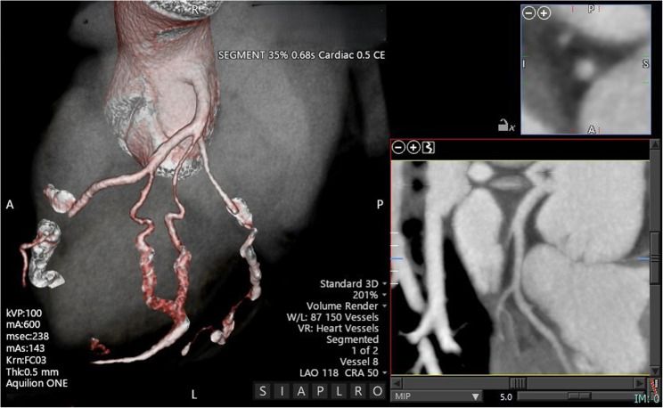
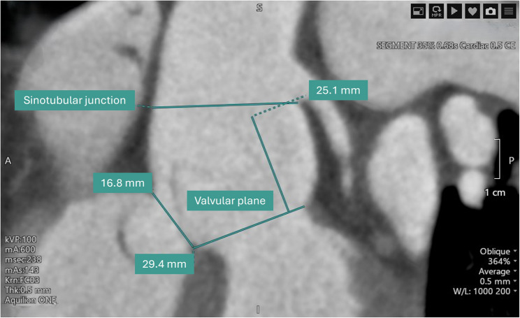
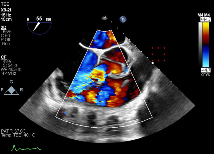
A 35-year-old male, without significant cardiovascular history, presented with recurrent palpitations. Initial echocardiographic evaluation demonstrated eccentric left ventricular hypertrophy, mild systolic dysfunction, suspicion of a ventricular septal defect, bicuspid aortic valve, and right ventricular dilation. Transesophageal echocardiography revealed an aneurysmal dilation of the right coronary sinus with an aorto-atrial/ventricular fistula, further confirmed by contrast-enhanced computed tomography angiography. Coronary angiography identified a single anomalous coronary artery with left dominance and absence of the right coronary artery. Surgical repair successfully closed the fistula, with mild post-operative aortic regurgitation. Follow-up at one year indicated normalization of cardiac dimensions and function, with stable moderate aortic valve regurgitation. Genetic sequencing found no identifiable mutations. Regular monitoring was recommended due to the potential risk of complications related to the bicuspid aortic valve and coronary anomaly.
期刊介绍:
Cardiovascular Ultrasound is an online journal, publishing peer-reviewed: original research; authoritative reviews; case reports on challenging and/or unusual diagnostic aspects; and expert opinions on new techniques and technologies. We are particularly interested in articles that include relevant images or video files, which provide an additional dimension to published articles and enhance understanding.
As an open access journal, Cardiovascular Ultrasound ensures high visibility for authors in addition to providing an up-to-date and freely available resource for the community. The journal welcomes discussion, and provides a forum for publishing opinion and debate ranging from biology to engineering to clinical echocardiography, with both speed and versatility.
 求助内容:
求助内容: 应助结果提醒方式:


