{"title":"快速改善的后部可逆性脑病综合征的影像学演变:1例报告。","authors":"Yilong He, Langtao He, Yetao Lin, Yitao He","doi":"10.4103/bc.bc_74_24","DOIUrl":null,"url":null,"abstract":"<p><p>Posterior reversible encephalopathy syndrome (PRES) is a rare neurological disorder with no specific clinical symptoms in the early stage; thus, early imaging identification is of great importance. A 29-year-old pregnant woman at 37 weeks experienced sudden generalized seizures accompanied by impaired consciousness. Brain computed tomography, conducted around 3 h after the onset, revealed symmetric areas of decreased density in the basal ganglia, brainstem, and suboccipital cortex, along with cerebral swelling. Following treatment with positive inotrope, diuretics, antihypertensives, and dehydration therapy, the patient regained clear consciousness on the 2<sup>nd</sup> day. On the 9<sup>th</sup> day postonset, a follow-up contrast-enhanced magnetic resonance imaging (MRI) showed slightly elevated signals in the bilateral occipital lobes on the T2 fluid-attenuated inversion recovery sequence. A subsequent brain MRI on day 47 postonset indicated no significant abnormal changes. Neuroimaging is pivotal for PRES diagnosis, revealing typical signs of widespread vasogenic edema in the posterior brain white matter, affecting the occipital lobes, cerebellum, brainstem, thalamus, and basal ganglia. With timely treatment, these lesions can partially or completely resolve within days or weeks.</p>","PeriodicalId":9288,"journal":{"name":"Brain Circulation","volume":"11 3","pages":"240-242"},"PeriodicalIF":4.8000,"publicationDate":"2025-03-04","publicationTypes":"Journal Article","fieldsOfStudy":null,"isOpenAccess":false,"openAccessPdf":"https://www.ncbi.nlm.nih.gov/pmc/articles/PMC12367258/pdf/","citationCount":"0","resultStr":"{\"title\":\"The evolving imaging of rapidly improving posterior reversible encephalopathy syndrome: A case report.\",\"authors\":\"Yilong He, Langtao He, Yetao Lin, Yitao He\",\"doi\":\"10.4103/bc.bc_74_24\",\"DOIUrl\":null,\"url\":null,\"abstract\":\"<p><p>Posterior reversible encephalopathy syndrome (PRES) is a rare neurological disorder with no specific clinical symptoms in the early stage; thus, early imaging identification is of great importance. A 29-year-old pregnant woman at 37 weeks experienced sudden generalized seizures accompanied by impaired consciousness. Brain computed tomography, conducted around 3 h after the onset, revealed symmetric areas of decreased density in the basal ganglia, brainstem, and suboccipital cortex, along with cerebral swelling. Following treatment with positive inotrope, diuretics, antihypertensives, and dehydration therapy, the patient regained clear consciousness on the 2<sup>nd</sup> day. On the 9<sup>th</sup> day postonset, a follow-up contrast-enhanced magnetic resonance imaging (MRI) showed slightly elevated signals in the bilateral occipital lobes on the T2 fluid-attenuated inversion recovery sequence. A subsequent brain MRI on day 47 postonset indicated no significant abnormal changes. Neuroimaging is pivotal for PRES diagnosis, revealing typical signs of widespread vasogenic edema in the posterior brain white matter, affecting the occipital lobes, cerebellum, brainstem, thalamus, and basal ganglia. With timely treatment, these lesions can partially or completely resolve within days or weeks.</p>\",\"PeriodicalId\":9288,\"journal\":{\"name\":\"Brain Circulation\",\"volume\":\"11 3\",\"pages\":\"240-242\"},\"PeriodicalIF\":4.8000,\"publicationDate\":\"2025-03-04\",\"publicationTypes\":\"Journal Article\",\"fieldsOfStudy\":null,\"isOpenAccess\":false,\"openAccessPdf\":\"https://www.ncbi.nlm.nih.gov/pmc/articles/PMC12367258/pdf/\",\"citationCount\":\"0\",\"resultStr\":null,\"platform\":\"Semanticscholar\",\"paperid\":null,\"PeriodicalName\":\"Brain Circulation\",\"FirstCategoryId\":\"3\",\"ListUrlMain\":\"https://doi.org/10.4103/bc.bc_74_24\",\"RegionNum\":4,\"RegionCategory\":\"医学\",\"ArticlePicture\":[],\"TitleCN\":null,\"AbstractTextCN\":null,\"PMCID\":null,\"EPubDate\":\"2025/7/1 0:00:00\",\"PubModel\":\"eCollection\",\"JCR\":\"Q3\",\"JCRName\":\"CLINICAL NEUROLOGY\",\"Score\":null,\"Total\":0}","platform":"Semanticscholar","paperid":null,"PeriodicalName":"Brain Circulation","FirstCategoryId":"3","ListUrlMain":"https://doi.org/10.4103/bc.bc_74_24","RegionNum":4,"RegionCategory":"医学","ArticlePicture":[],"TitleCN":null,"AbstractTextCN":null,"PMCID":null,"EPubDate":"2025/7/1 0:00:00","PubModel":"eCollection","JCR":"Q3","JCRName":"CLINICAL NEUROLOGY","Score":null,"Total":0}
The evolving imaging of rapidly improving posterior reversible encephalopathy syndrome: A case report.
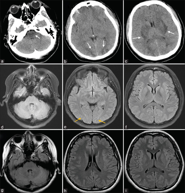
Posterior reversible encephalopathy syndrome (PRES) is a rare neurological disorder with no specific clinical symptoms in the early stage; thus, early imaging identification is of great importance. A 29-year-old pregnant woman at 37 weeks experienced sudden generalized seizures accompanied by impaired consciousness. Brain computed tomography, conducted around 3 h after the onset, revealed symmetric areas of decreased density in the basal ganglia, brainstem, and suboccipital cortex, along with cerebral swelling. Following treatment with positive inotrope, diuretics, antihypertensives, and dehydration therapy, the patient regained clear consciousness on the 2nd day. On the 9th day postonset, a follow-up contrast-enhanced magnetic resonance imaging (MRI) showed slightly elevated signals in the bilateral occipital lobes on the T2 fluid-attenuated inversion recovery sequence. A subsequent brain MRI on day 47 postonset indicated no significant abnormal changes. Neuroimaging is pivotal for PRES diagnosis, revealing typical signs of widespread vasogenic edema in the posterior brain white matter, affecting the occipital lobes, cerebellum, brainstem, thalamus, and basal ganglia. With timely treatment, these lesions can partially or completely resolve within days or weeks.

 求助内容:
求助内容: 应助结果提醒方式:


