Beatriz de Lucena Ribeiro, Ana Lucia Passos Peixoto, Ana Paula Couto, Rafael Erthal Robbs, Wander Borges, Julieta Micherif, Giovanna Provenzano, Raul N G Vianna
{"title":"单侧视网膜色素上皮发育不良(URPED)患者的显微视野和多焦视网膜电图。","authors":"Beatriz de Lucena Ribeiro, Ana Lucia Passos Peixoto, Ana Paula Couto, Rafael Erthal Robbs, Wander Borges, Julieta Micherif, Giovanna Provenzano, Raul N G Vianna","doi":"10.1155/crop/7911612","DOIUrl":null,"url":null,"abstract":"<p><p><b>Introduction:</b> This study is aimed at describing a patient with unilateral retinal pigment epithelium dysgenesis (URPED) using multimodal retinal imaging combined with ocular microperimetry and multifocal electroretinogram (ERG) analysis. <b>Case presentation:</b> A 56-year-old healthy male was referred for a routine ophthalmologic control. His best corrected visual acuity was 20/20 and 20/25 in the right and left eye, respectively. Fundus examination of the left eye revealed a well-circumscribed, large yellowish-white lesion on the posterior pole that extended from the peripapillary region to the inferior temporal vascular arcade, sparing the fovea. This characteristic fundus picture led us to the diagnosis of URPED. Microperimetry demonstrated a progressive decrease of sensitivity from the normal retina toward the lesion borders, reaching a value of 0 dB at its center. Multifocal ERG displayed a reduction of central amplitudes in the involved eye. <b>Discussion:</b> Our findings indicate a varied degree of sensitivity at the site of the lesion. Despite good visual acuity, multifocal ERG revealed reduced macular function.</p>","PeriodicalId":9603,"journal":{"name":"Case Reports in Ophthalmological Medicine","volume":"2025 ","pages":"7911612"},"PeriodicalIF":0.4000,"publicationDate":"2025-08-14","publicationTypes":"Journal Article","fieldsOfStudy":null,"isOpenAccess":false,"openAccessPdf":"https://www.ncbi.nlm.nih.gov/pmc/articles/PMC12370388/pdf/","citationCount":"0","resultStr":"{\"title\":\"Microperimetry and Multifocal Electroretinogram in a Patient With Unilateral Retinal Pigment Epithelium Dysgenesis (URPED).\",\"authors\":\"Beatriz de Lucena Ribeiro, Ana Lucia Passos Peixoto, Ana Paula Couto, Rafael Erthal Robbs, Wander Borges, Julieta Micherif, Giovanna Provenzano, Raul N G Vianna\",\"doi\":\"10.1155/crop/7911612\",\"DOIUrl\":null,\"url\":null,\"abstract\":\"<p><p><b>Introduction:</b> This study is aimed at describing a patient with unilateral retinal pigment epithelium dysgenesis (URPED) using multimodal retinal imaging combined with ocular microperimetry and multifocal electroretinogram (ERG) analysis. <b>Case presentation:</b> A 56-year-old healthy male was referred for a routine ophthalmologic control. His best corrected visual acuity was 20/20 and 20/25 in the right and left eye, respectively. Fundus examination of the left eye revealed a well-circumscribed, large yellowish-white lesion on the posterior pole that extended from the peripapillary region to the inferior temporal vascular arcade, sparing the fovea. This characteristic fundus picture led us to the diagnosis of URPED. Microperimetry demonstrated a progressive decrease of sensitivity from the normal retina toward the lesion borders, reaching a value of 0 dB at its center. Multifocal ERG displayed a reduction of central amplitudes in the involved eye. <b>Discussion:</b> Our findings indicate a varied degree of sensitivity at the site of the lesion. Despite good visual acuity, multifocal ERG revealed reduced macular function.</p>\",\"PeriodicalId\":9603,\"journal\":{\"name\":\"Case Reports in Ophthalmological Medicine\",\"volume\":\"2025 \",\"pages\":\"7911612\"},\"PeriodicalIF\":0.4000,\"publicationDate\":\"2025-08-14\",\"publicationTypes\":\"Journal Article\",\"fieldsOfStudy\":null,\"isOpenAccess\":false,\"openAccessPdf\":\"https://www.ncbi.nlm.nih.gov/pmc/articles/PMC12370388/pdf/\",\"citationCount\":\"0\",\"resultStr\":null,\"platform\":\"Semanticscholar\",\"paperid\":null,\"PeriodicalName\":\"Case Reports in Ophthalmological Medicine\",\"FirstCategoryId\":\"1085\",\"ListUrlMain\":\"https://doi.org/10.1155/crop/7911612\",\"RegionNum\":0,\"RegionCategory\":null,\"ArticlePicture\":[],\"TitleCN\":null,\"AbstractTextCN\":null,\"PMCID\":null,\"EPubDate\":\"2025/1/1 0:00:00\",\"PubModel\":\"eCollection\",\"JCR\":\"Q4\",\"JCRName\":\"OPHTHALMOLOGY\",\"Score\":null,\"Total\":0}","platform":"Semanticscholar","paperid":null,"PeriodicalName":"Case Reports in Ophthalmological Medicine","FirstCategoryId":"1085","ListUrlMain":"https://doi.org/10.1155/crop/7911612","RegionNum":0,"RegionCategory":null,"ArticlePicture":[],"TitleCN":null,"AbstractTextCN":null,"PMCID":null,"EPubDate":"2025/1/1 0:00:00","PubModel":"eCollection","JCR":"Q4","JCRName":"OPHTHALMOLOGY","Score":null,"Total":0}
Microperimetry and Multifocal Electroretinogram in a Patient With Unilateral Retinal Pigment Epithelium Dysgenesis (URPED).
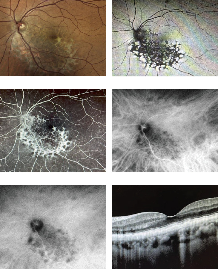
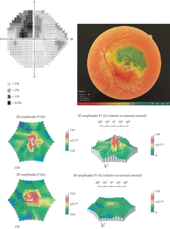
Introduction: This study is aimed at describing a patient with unilateral retinal pigment epithelium dysgenesis (URPED) using multimodal retinal imaging combined with ocular microperimetry and multifocal electroretinogram (ERG) analysis. Case presentation: A 56-year-old healthy male was referred for a routine ophthalmologic control. His best corrected visual acuity was 20/20 and 20/25 in the right and left eye, respectively. Fundus examination of the left eye revealed a well-circumscribed, large yellowish-white lesion on the posterior pole that extended from the peripapillary region to the inferior temporal vascular arcade, sparing the fovea. This characteristic fundus picture led us to the diagnosis of URPED. Microperimetry demonstrated a progressive decrease of sensitivity from the normal retina toward the lesion borders, reaching a value of 0 dB at its center. Multifocal ERG displayed a reduction of central amplitudes in the involved eye. Discussion: Our findings indicate a varied degree of sensitivity at the site of the lesion. Despite good visual acuity, multifocal ERG revealed reduced macular function.

 求助内容:
求助内容: 应助结果提醒方式:


