{"title":"急性真菌性白内障后眼内炎在眼内炎治疗中的研究:EMS报告","authors":"Taraprasad Das, Akash Belenje, Joveeta Joseph, Suchita Pandey, Dhanush Pandya, Rudvij Pandya, Umesh Chandra Behera, Vivek Pravin Dave","doi":"10.1136/bmjophth-2025-002190","DOIUrl":null,"url":null,"abstract":"<p><strong>Aim: </strong>Investigate and characterise acute post-cataract fungal endophthalmitis pooled from the Endophthalmitis Management Study occurring within 6 weeks of primary surgery.</p><p><strong>Methods: </strong>The fungal infection was confirmed through conventional and molecular microbiology work-ups and antifungal susceptibility testing. Clinical examination included measurement of distant vision and intraocular pressure, anterior segment photo documentation and inflammation score (IS) measurement. Per the microbiology report, the eyes were divided into culture-positive (C+), sequencing-positive (S+) and sequencing-positive-unidentified (U+) fungi. Clinical correlations and statistical comparisons were performed between these three cohorts.</p><p><strong>Results: </strong>The study identified 21 patients with fungal endophthalmitis; it was 9.5% (21 of 220) of all acute post-cataract endophthalmitis in this study. Per the microbiology report, C+, S+ and U+ were 6, 9 and 6 patients, respectively. <i>Fusarium</i> and <i>Aspergillus</i> spp were the common fungi. The C+ fungi had higher presenting IS (p=0.023), shorter time to symptoms, worse presenting vision, corneal abscess (p=0.030) and higher probability of repeat intervention (p=0.042) than the other two groups. In the C+ group, the final vision of >20/400 was less (p=0.046) and phthisis bulbi was higher (p=0.010). All culturable fungi were resistant to amphotericin B and voriconazole.</p><p><strong>Conclusion: </strong>There is a 10% probability of acute post-cataract fungal endophthalmitis in India. The eyes presenting with corneal abscesses carry a higher risk. The polymicrobial infections shown in this cohort should be interpreted cautiously since next-generation sequencing detects DNA from all organisms, including residual or low-abundance or non-viable organisms that traditional culture might miss. Despite this, the new molecular microbiology technology is necessary to confirm diagnosis and expedite appropriate treatment. Given multi-antifungal agent resistance, routine susceptibility testing must be considered.</p>","PeriodicalId":9286,"journal":{"name":"BMJ Open Ophthalmology","volume":"10 1","pages":""},"PeriodicalIF":2.2000,"publicationDate":"2025-08-19","publicationTypes":"Journal Article","fieldsOfStudy":null,"isOpenAccess":false,"openAccessPdf":"https://www.ncbi.nlm.nih.gov/pmc/articles/PMC12366611/pdf/","citationCount":"0","resultStr":"{\"title\":\"Acute fungal post-cataract endophthalmitis in the endophthalmitis management study: EMS report 7.\",\"authors\":\"Taraprasad Das, Akash Belenje, Joveeta Joseph, Suchita Pandey, Dhanush Pandya, Rudvij Pandya, Umesh Chandra Behera, Vivek Pravin Dave\",\"doi\":\"10.1136/bmjophth-2025-002190\",\"DOIUrl\":null,\"url\":null,\"abstract\":\"<p><strong>Aim: </strong>Investigate and characterise acute post-cataract fungal endophthalmitis pooled from the Endophthalmitis Management Study occurring within 6 weeks of primary surgery.</p><p><strong>Methods: </strong>The fungal infection was confirmed through conventional and molecular microbiology work-ups and antifungal susceptibility testing. Clinical examination included measurement of distant vision and intraocular pressure, anterior segment photo documentation and inflammation score (IS) measurement. Per the microbiology report, the eyes were divided into culture-positive (C+), sequencing-positive (S+) and sequencing-positive-unidentified (U+) fungi. Clinical correlations and statistical comparisons were performed between these three cohorts.</p><p><strong>Results: </strong>The study identified 21 patients with fungal endophthalmitis; it was 9.5% (21 of 220) of all acute post-cataract endophthalmitis in this study. Per the microbiology report, C+, S+ and U+ were 6, 9 and 6 patients, respectively. <i>Fusarium</i> and <i>Aspergillus</i> spp were the common fungi. The C+ fungi had higher presenting IS (p=0.023), shorter time to symptoms, worse presenting vision, corneal abscess (p=0.030) and higher probability of repeat intervention (p=0.042) than the other two groups. In the C+ group, the final vision of >20/400 was less (p=0.046) and phthisis bulbi was higher (p=0.010). All culturable fungi were resistant to amphotericin B and voriconazole.</p><p><strong>Conclusion: </strong>There is a 10% probability of acute post-cataract fungal endophthalmitis in India. The eyes presenting with corneal abscesses carry a higher risk. The polymicrobial infections shown in this cohort should be interpreted cautiously since next-generation sequencing detects DNA from all organisms, including residual or low-abundance or non-viable organisms that traditional culture might miss. Despite this, the new molecular microbiology technology is necessary to confirm diagnosis and expedite appropriate treatment. Given multi-antifungal agent resistance, routine susceptibility testing must be considered.</p>\",\"PeriodicalId\":9286,\"journal\":{\"name\":\"BMJ Open Ophthalmology\",\"volume\":\"10 1\",\"pages\":\"\"},\"PeriodicalIF\":2.2000,\"publicationDate\":\"2025-08-19\",\"publicationTypes\":\"Journal Article\",\"fieldsOfStudy\":null,\"isOpenAccess\":false,\"openAccessPdf\":\"https://www.ncbi.nlm.nih.gov/pmc/articles/PMC12366611/pdf/\",\"citationCount\":\"0\",\"resultStr\":null,\"platform\":\"Semanticscholar\",\"paperid\":null,\"PeriodicalName\":\"BMJ Open Ophthalmology\",\"FirstCategoryId\":\"1085\",\"ListUrlMain\":\"https://doi.org/10.1136/bmjophth-2025-002190\",\"RegionNum\":0,\"RegionCategory\":null,\"ArticlePicture\":[],\"TitleCN\":null,\"AbstractTextCN\":null,\"PMCID\":null,\"EPubDate\":\"\",\"PubModel\":\"\",\"JCR\":\"Q2\",\"JCRName\":\"OPHTHALMOLOGY\",\"Score\":null,\"Total\":0}","platform":"Semanticscholar","paperid":null,"PeriodicalName":"BMJ Open Ophthalmology","FirstCategoryId":"1085","ListUrlMain":"https://doi.org/10.1136/bmjophth-2025-002190","RegionNum":0,"RegionCategory":null,"ArticlePicture":[],"TitleCN":null,"AbstractTextCN":null,"PMCID":null,"EPubDate":"","PubModel":"","JCR":"Q2","JCRName":"OPHTHALMOLOGY","Score":null,"Total":0}
Acute fungal post-cataract endophthalmitis in the endophthalmitis management study: EMS report 7.
Aim: Investigate and characterise acute post-cataract fungal endophthalmitis pooled from the Endophthalmitis Management Study occurring within 6 weeks of primary surgery.
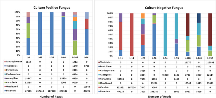
Methods: The fungal infection was confirmed through conventional and molecular microbiology work-ups and antifungal susceptibility testing. Clinical examination included measurement of distant vision and intraocular pressure, anterior segment photo documentation and inflammation score (IS) measurement. Per the microbiology report, the eyes were divided into culture-positive (C+), sequencing-positive (S+) and sequencing-positive-unidentified (U+) fungi. Clinical correlations and statistical comparisons were performed between these three cohorts.
Results: The study identified 21 patients with fungal endophthalmitis; it was 9.5% (21 of 220) of all acute post-cataract endophthalmitis in this study. Per the microbiology report, C+, S+ and U+ were 6, 9 and 6 patients, respectively. Fusarium and Aspergillus spp were the common fungi. The C+ fungi had higher presenting IS (p=0.023), shorter time to symptoms, worse presenting vision, corneal abscess (p=0.030) and higher probability of repeat intervention (p=0.042) than the other two groups. In the C+ group, the final vision of >20/400 was less (p=0.046) and phthisis bulbi was higher (p=0.010). All culturable fungi were resistant to amphotericin B and voriconazole.
Conclusion: There is a 10% probability of acute post-cataract fungal endophthalmitis in India. The eyes presenting with corneal abscesses carry a higher risk. The polymicrobial infections shown in this cohort should be interpreted cautiously since next-generation sequencing detects DNA from all organisms, including residual or low-abundance or non-viable organisms that traditional culture might miss. Despite this, the new molecular microbiology technology is necessary to confirm diagnosis and expedite appropriate treatment. Given multi-antifungal agent resistance, routine susceptibility testing must be considered.

 求助内容:
求助内容: 应助结果提醒方式:


