{"title":"眼科基础模型:超广角眼底图像人工智能辅助诊断近视黄斑病和后葡萄肿的初步研究。","authors":"Juzhao Zhang, Tao Yu, Mengjia Zhang, Yuzhu Zhang, Yingyan Ma, Wenwen Xue, Hao Zhou, Senlin Lin, Haidong Zou, Xian Xu","doi":"10.1136/bmjophth-2024-002073","DOIUrl":null,"url":null,"abstract":"<p><strong>Objectives: </strong>This study aims to detect characteristic fundus changes in pathological myopia using deep learning (DL)-based analysis of ultra-widefield (UWF) fundus imaging.</p><p><strong>Methods: </strong>Following the exclusion of low-quality images, this cross-sectional study used 1105 UWF images from 543 patients with high myopia to develop the model, along with 293 images from 150 patients with high myopia for external testing. All images were retrospectively collected from patients with high myopia at Shanghai General Hospital and Shanghai Eye Diseases Prevention and Treatment Center between 2018 and 2024. We trained a DL model based on an ophthalmology foundational model to detect myopic maculopathy (MM) and posterior staphyloma (PS).</p><p><strong>Results: </strong>The proposed RETFound-enhanced model demonstrated robust performance. For five-category classification of MM, it achieved 65.4% accuracy and an F1 score of 0.648, outperforming other methods. In three-category MM classification, it achieved 79.4% accuracy and an F1 score of 0.793. For PS detection, the model reached 84.1% accuracy, an F1 score of 0.814 and an area under the receiver operating characteristic curve (AUROC) of 0.886, highlighting its effectiveness as a screening tool. External validation showed consistent performance, with 64.4% accuracy for five-category MM classification, 79.8% accuracy for three-category classification and 81.2% accuracy for PS, confirming robustness across cohorts.</p><p><strong>Conclusions: </strong>This study presents an effective diagnostic model for pathological myopia using UWF fundus imaging and a foundation model. The integration of DL with non-mydriatic UWF fundus imaging demonstrates promising potential for applications in primary healthcare, particularly in underserved areas, enabling accessible screening for high myopia-related fundus changes.</p>","PeriodicalId":9286,"journal":{"name":"BMJ Open Ophthalmology","volume":"10 1","pages":""},"PeriodicalIF":2.2000,"publicationDate":"2025-08-28","publicationTypes":"Journal Article","fieldsOfStudy":null,"isOpenAccess":false,"openAccessPdf":"https://www.ncbi.nlm.nih.gov/pmc/articles/PMC12410655/pdf/","citationCount":"0","resultStr":"{\"title\":\"Foundation models in ophthalmology: a preliminary study on AI-assisted diagnosis of myopic maculopathy and posterior staphyloma using ultra-widefield fundus images.\",\"authors\":\"Juzhao Zhang, Tao Yu, Mengjia Zhang, Yuzhu Zhang, Yingyan Ma, Wenwen Xue, Hao Zhou, Senlin Lin, Haidong Zou, Xian Xu\",\"doi\":\"10.1136/bmjophth-2024-002073\",\"DOIUrl\":null,\"url\":null,\"abstract\":\"<p><strong>Objectives: </strong>This study aims to detect characteristic fundus changes in pathological myopia using deep learning (DL)-based analysis of ultra-widefield (UWF) fundus imaging.</p><p><strong>Methods: </strong>Following the exclusion of low-quality images, this cross-sectional study used 1105 UWF images from 543 patients with high myopia to develop the model, along with 293 images from 150 patients with high myopia for external testing. All images were retrospectively collected from patients with high myopia at Shanghai General Hospital and Shanghai Eye Diseases Prevention and Treatment Center between 2018 and 2024. We trained a DL model based on an ophthalmology foundational model to detect myopic maculopathy (MM) and posterior staphyloma (PS).</p><p><strong>Results: </strong>The proposed RETFound-enhanced model demonstrated robust performance. For five-category classification of MM, it achieved 65.4% accuracy and an F1 score of 0.648, outperforming other methods. In three-category MM classification, it achieved 79.4% accuracy and an F1 score of 0.793. For PS detection, the model reached 84.1% accuracy, an F1 score of 0.814 and an area under the receiver operating characteristic curve (AUROC) of 0.886, highlighting its effectiveness as a screening tool. External validation showed consistent performance, with 64.4% accuracy for five-category MM classification, 79.8% accuracy for three-category classification and 81.2% accuracy for PS, confirming robustness across cohorts.</p><p><strong>Conclusions: </strong>This study presents an effective diagnostic model for pathological myopia using UWF fundus imaging and a foundation model. The integration of DL with non-mydriatic UWF fundus imaging demonstrates promising potential for applications in primary healthcare, particularly in underserved areas, enabling accessible screening for high myopia-related fundus changes.</p>\",\"PeriodicalId\":9286,\"journal\":{\"name\":\"BMJ Open Ophthalmology\",\"volume\":\"10 1\",\"pages\":\"\"},\"PeriodicalIF\":2.2000,\"publicationDate\":\"2025-08-28\",\"publicationTypes\":\"Journal Article\",\"fieldsOfStudy\":null,\"isOpenAccess\":false,\"openAccessPdf\":\"https://www.ncbi.nlm.nih.gov/pmc/articles/PMC12410655/pdf/\",\"citationCount\":\"0\",\"resultStr\":null,\"platform\":\"Semanticscholar\",\"paperid\":null,\"PeriodicalName\":\"BMJ Open Ophthalmology\",\"FirstCategoryId\":\"1085\",\"ListUrlMain\":\"https://doi.org/10.1136/bmjophth-2024-002073\",\"RegionNum\":0,\"RegionCategory\":null,\"ArticlePicture\":[],\"TitleCN\":null,\"AbstractTextCN\":null,\"PMCID\":null,\"EPubDate\":\"\",\"PubModel\":\"\",\"JCR\":\"Q2\",\"JCRName\":\"OPHTHALMOLOGY\",\"Score\":null,\"Total\":0}","platform":"Semanticscholar","paperid":null,"PeriodicalName":"BMJ Open Ophthalmology","FirstCategoryId":"1085","ListUrlMain":"https://doi.org/10.1136/bmjophth-2024-002073","RegionNum":0,"RegionCategory":null,"ArticlePicture":[],"TitleCN":null,"AbstractTextCN":null,"PMCID":null,"EPubDate":"","PubModel":"","JCR":"Q2","JCRName":"OPHTHALMOLOGY","Score":null,"Total":0}
Foundation models in ophthalmology: a preliminary study on AI-assisted diagnosis of myopic maculopathy and posterior staphyloma using ultra-widefield fundus images.
Objectives: This study aims to detect characteristic fundus changes in pathological myopia using deep learning (DL)-based analysis of ultra-widefield (UWF) fundus imaging.
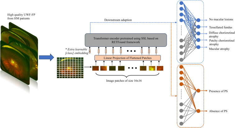
Methods: Following the exclusion of low-quality images, this cross-sectional study used 1105 UWF images from 543 patients with high myopia to develop the model, along with 293 images from 150 patients with high myopia for external testing. All images were retrospectively collected from patients with high myopia at Shanghai General Hospital and Shanghai Eye Diseases Prevention and Treatment Center between 2018 and 2024. We trained a DL model based on an ophthalmology foundational model to detect myopic maculopathy (MM) and posterior staphyloma (PS).
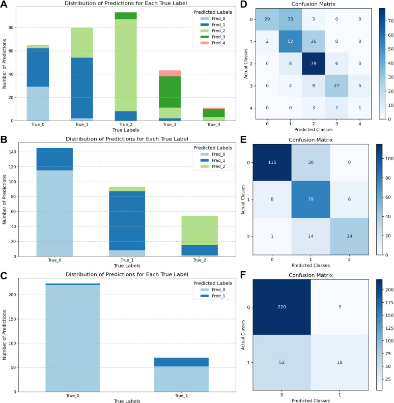
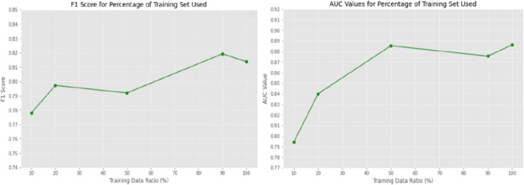
Results: The proposed RETFound-enhanced model demonstrated robust performance. For five-category classification of MM, it achieved 65.4% accuracy and an F1 score of 0.648, outperforming other methods. In three-category MM classification, it achieved 79.4% accuracy and an F1 score of 0.793. For PS detection, the model reached 84.1% accuracy, an F1 score of 0.814 and an area under the receiver operating characteristic curve (AUROC) of 0.886, highlighting its effectiveness as a screening tool. External validation showed consistent performance, with 64.4% accuracy for five-category MM classification, 79.8% accuracy for three-category classification and 81.2% accuracy for PS, confirming robustness across cohorts.
Conclusions: This study presents an effective diagnostic model for pathological myopia using UWF fundus imaging and a foundation model. The integration of DL with non-mydriatic UWF fundus imaging demonstrates promising potential for applications in primary healthcare, particularly in underserved areas, enabling accessible screening for high myopia-related fundus changes.

 求助内容:
求助内容: 应助结果提醒方式:


