Francis Letendre, Abigail Blackburn, Michael Twardowski
{"title":"鞭毛藻中机械刺激生物发光的峰值强度与细胞表面积的联系。","authors":"Francis Letendre, Abigail Blackburn, Michael Twardowski","doi":"10.1242/bio.062190","DOIUrl":null,"url":null,"abstract":"<p><p>Mechanically stimulated bioluminescence (MSL) is present in most planktonic clades and marine ecosystems. The first flash kinetic parameters (FFKPs) and spectral properties are often species specific, making MSL a powerful tool for in situ ID and biodiversity assessments. The peak intensity (PI) of mechanically stimulated bioluminescence was measured for five species of dinoflagellates: Alexandrium monilatum, Lingulodinium polyedra, Pyrocystis fusiformis, Pyrocystis noctiluca and Pyrodinium bahamense. Peak intensity was assessed with respect to organism cell surface area and volume, building upon Seliger's rule, where previously a relationship was found linking cell surface area and total mechanically stimulated light (TMSL) ( Buskey and Swift, 1990). These dinoflagellate species were chosen to cover a wide range of peak intensities (108 to 1010 photons/s) and surface area (104 to 106μm2). Individual cells were isolated and individually photographed under a compound microscope, where cell size was measured. They were then dark-adapted and first flash emission from mechanical stimulation was measured with the Underwater Bioluminescence Assessment Tool (UBAT) from Seabird Scientific (www.seabird.com). Distributions of PI and surface area across all species were compared using non-parametric ANOVAs and a linear regression model, uncovering a strong positive correlation and strength of fit across all species between peak intensity and both cell surface area and volume. This study provides insight into understanding and potentially predicting the bioluminescence of organisms often responsible for significant primary and secondary productivity in marine waters with subsequent global impacts on fisheries and ecology. Bioluminescence measurements may also be a powerful tool for understanding plankton composition, ecology, and diversity.</p>","PeriodicalId":9216,"journal":{"name":"Biology Open","volume":" ","pages":""},"PeriodicalIF":1.7000,"publicationDate":"2025-09-15","publicationTypes":"Journal Article","fieldsOfStudy":null,"isOpenAccess":false,"openAccessPdf":"https://www.ncbi.nlm.nih.gov/pmc/articles/PMC12486207/pdf/","citationCount":"0","resultStr":"{\"title\":\"Linking peak intensity of mechanically stimulated bioluminescence and cell surface area in dinoflagellates.\",\"authors\":\"Francis Letendre, Abigail Blackburn, Michael Twardowski\",\"doi\":\"10.1242/bio.062190\",\"DOIUrl\":null,\"url\":null,\"abstract\":\"<p><p>Mechanically stimulated bioluminescence (MSL) is present in most planktonic clades and marine ecosystems. The first flash kinetic parameters (FFKPs) and spectral properties are often species specific, making MSL a powerful tool for in situ ID and biodiversity assessments. The peak intensity (PI) of mechanically stimulated bioluminescence was measured for five species of dinoflagellates: Alexandrium monilatum, Lingulodinium polyedra, Pyrocystis fusiformis, Pyrocystis noctiluca and Pyrodinium bahamense. Peak intensity was assessed with respect to organism cell surface area and volume, building upon Seliger's rule, where previously a relationship was found linking cell surface area and total mechanically stimulated light (TMSL) ( Buskey and Swift, 1990). These dinoflagellate species were chosen to cover a wide range of peak intensities (108 to 1010 photons/s) and surface area (104 to 106μm2). Individual cells were isolated and individually photographed under a compound microscope, where cell size was measured. They were then dark-adapted and first flash emission from mechanical stimulation was measured with the Underwater Bioluminescence Assessment Tool (UBAT) from Seabird Scientific (www.seabird.com). Distributions of PI and surface area across all species were compared using non-parametric ANOVAs and a linear regression model, uncovering a strong positive correlation and strength of fit across all species between peak intensity and both cell surface area and volume. This study provides insight into understanding and potentially predicting the bioluminescence of organisms often responsible for significant primary and secondary productivity in marine waters with subsequent global impacts on fisheries and ecology. Bioluminescence measurements may also be a powerful tool for understanding plankton composition, ecology, and diversity.</p>\",\"PeriodicalId\":9216,\"journal\":{\"name\":\"Biology Open\",\"volume\":\" \",\"pages\":\"\"},\"PeriodicalIF\":1.7000,\"publicationDate\":\"2025-09-15\",\"publicationTypes\":\"Journal Article\",\"fieldsOfStudy\":null,\"isOpenAccess\":false,\"openAccessPdf\":\"https://www.ncbi.nlm.nih.gov/pmc/articles/PMC12486207/pdf/\",\"citationCount\":\"0\",\"resultStr\":null,\"platform\":\"Semanticscholar\",\"paperid\":null,\"PeriodicalName\":\"Biology Open\",\"FirstCategoryId\":\"99\",\"ListUrlMain\":\"https://doi.org/10.1242/bio.062190\",\"RegionNum\":4,\"RegionCategory\":\"生物学\",\"ArticlePicture\":[],\"TitleCN\":null,\"AbstractTextCN\":null,\"PMCID\":null,\"EPubDate\":\"2025/9/19 0:00:00\",\"PubModel\":\"Epub\",\"JCR\":\"Q3\",\"JCRName\":\"BIOLOGY\",\"Score\":null,\"Total\":0}","platform":"Semanticscholar","paperid":null,"PeriodicalName":"Biology Open","FirstCategoryId":"99","ListUrlMain":"https://doi.org/10.1242/bio.062190","RegionNum":4,"RegionCategory":"生物学","ArticlePicture":[],"TitleCN":null,"AbstractTextCN":null,"PMCID":null,"EPubDate":"2025/9/19 0:00:00","PubModel":"Epub","JCR":"Q3","JCRName":"BIOLOGY","Score":null,"Total":0}
Linking peak intensity of mechanically stimulated bioluminescence and cell surface area in dinoflagellates.
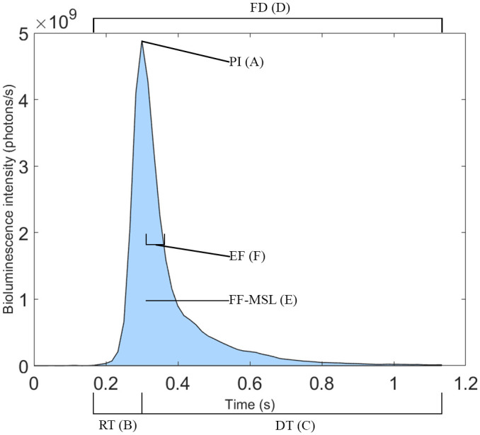
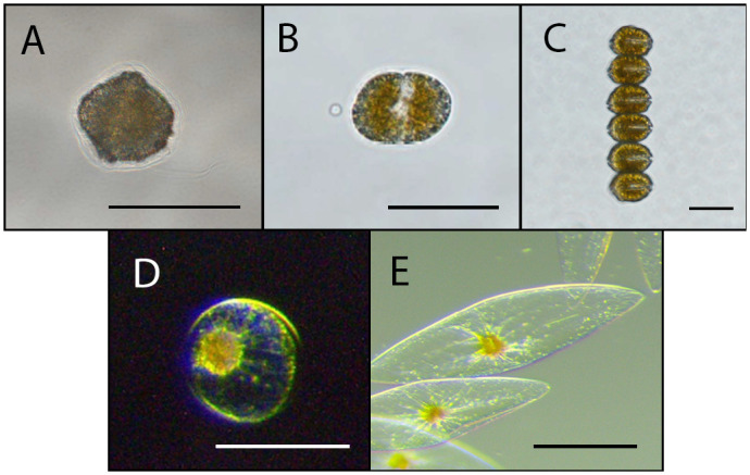
Mechanically stimulated bioluminescence (MSL) is present in most planktonic clades and marine ecosystems. The first flash kinetic parameters (FFKPs) and spectral properties are often species specific, making MSL a powerful tool for in situ ID and biodiversity assessments. The peak intensity (PI) of mechanically stimulated bioluminescence was measured for five species of dinoflagellates: Alexandrium monilatum, Lingulodinium polyedra, Pyrocystis fusiformis, Pyrocystis noctiluca and Pyrodinium bahamense. Peak intensity was assessed with respect to organism cell surface area and volume, building upon Seliger's rule, where previously a relationship was found linking cell surface area and total mechanically stimulated light (TMSL) ( Buskey and Swift, 1990). These dinoflagellate species were chosen to cover a wide range of peak intensities (108 to 1010 photons/s) and surface area (104 to 106μm2). Individual cells were isolated and individually photographed under a compound microscope, where cell size was measured. They were then dark-adapted and first flash emission from mechanical stimulation was measured with the Underwater Bioluminescence Assessment Tool (UBAT) from Seabird Scientific (www.seabird.com). Distributions of PI and surface area across all species were compared using non-parametric ANOVAs and a linear regression model, uncovering a strong positive correlation and strength of fit across all species between peak intensity and both cell surface area and volume. This study provides insight into understanding and potentially predicting the bioluminescence of organisms often responsible for significant primary and secondary productivity in marine waters with subsequent global impacts on fisheries and ecology. Bioluminescence measurements may also be a powerful tool for understanding plankton composition, ecology, and diversity.
期刊介绍:
Biology Open (BiO) is an online Open Access journal that publishes peer-reviewed original research across all aspects of the biological sciences. BiO aims to provide rapid publication for scientifically sound observations and valid conclusions, without a requirement for perceived impact.
 求助内容:
求助内容: 应助结果提醒方式:


