Elibeth Monroy, Leonardo Aguilar-Hernandez, Fidel de la Cruz-López, Gonzalo Flores, Julio César Morales-Medina
{"title":"雄性小鼠树突状脊柱退化与年龄相关的识别和空间记忆衰退有关。","authors":"Elibeth Monroy, Leonardo Aguilar-Hernandez, Fidel de la Cruz-López, Gonzalo Flores, Julio César Morales-Medina","doi":"10.1007/s00429-025-03002-7","DOIUrl":null,"url":null,"abstract":"<p><p>Human populations are experiencing an increase in aging, which is associated with cognitive deficits. Animal models of aging have shown that these behavioral impairments are associated with neuroarchitecture modifications in the prefrontal cortex (PFC) and hippocampus; however, most studies have focused on rats or lack multiple key ages. In this study, we evaluated spatial and recognition memory in male mice at critical ages [3 months (M), 6, 12 and 18] using the Morris water maze (MWM) and novel object recognition test (NORT), respectively. Moreover, we quantified dendritic arborization, spine density and the type of spines in the PFC, CA1 hippocampus and nucleus Accumbens Core (NAcC). Locomotion, assessed in the first phase of NORT, revealed age-dependent reductions. Notably, the 18 M group revealed significant recognition memory deficits. Spatial memory impairments were especially evident at the 12 M group in the MWM. Spine density was increased at 6 M in the NAcC, whereas a reduction was noted at 12 M and 18 M in the PFC. Morphological assessment of spines indicated age-dependent changes, including a notable increase in the proportion of thin spines in the CA1 and PFC regions. However, dendritic arborization remained largely unchanged across the examined brain regions and age groups. Overall, our findings observed age-dependent alterations in memory and morphological alterations in spines in mice, emerging as possible contributors to cognitive decline. These results highlight the potential for anti-aging interventions targeting synaptic structures to enhance cognitive health and extend the healthspan of aging individuals.</p>","PeriodicalId":9145,"journal":{"name":"Brain Structure & Function","volume":"230 7","pages":"142"},"PeriodicalIF":2.9000,"publicationDate":"2025-08-28","publicationTypes":"Journal Article","fieldsOfStudy":null,"isOpenAccess":false,"openAccessPdf":"https://www.ncbi.nlm.nih.gov/pmc/articles/PMC12394372/pdf/","citationCount":"0","resultStr":"{\"title\":\"Dendritic spine degeneration is associated with age-related decline in recognition and spatial memory in male mice.\",\"authors\":\"Elibeth Monroy, Leonardo Aguilar-Hernandez, Fidel de la Cruz-López, Gonzalo Flores, Julio César Morales-Medina\",\"doi\":\"10.1007/s00429-025-03002-7\",\"DOIUrl\":null,\"url\":null,\"abstract\":\"<p><p>Human populations are experiencing an increase in aging, which is associated with cognitive deficits. Animal models of aging have shown that these behavioral impairments are associated with neuroarchitecture modifications in the prefrontal cortex (PFC) and hippocampus; however, most studies have focused on rats or lack multiple key ages. In this study, we evaluated spatial and recognition memory in male mice at critical ages [3 months (M), 6, 12 and 18] using the Morris water maze (MWM) and novel object recognition test (NORT), respectively. Moreover, we quantified dendritic arborization, spine density and the type of spines in the PFC, CA1 hippocampus and nucleus Accumbens Core (NAcC). Locomotion, assessed in the first phase of NORT, revealed age-dependent reductions. Notably, the 18 M group revealed significant recognition memory deficits. Spatial memory impairments were especially evident at the 12 M group in the MWM. Spine density was increased at 6 M in the NAcC, whereas a reduction was noted at 12 M and 18 M in the PFC. Morphological assessment of spines indicated age-dependent changes, including a notable increase in the proportion of thin spines in the CA1 and PFC regions. However, dendritic arborization remained largely unchanged across the examined brain regions and age groups. Overall, our findings observed age-dependent alterations in memory and morphological alterations in spines in mice, emerging as possible contributors to cognitive decline. These results highlight the potential for anti-aging interventions targeting synaptic structures to enhance cognitive health and extend the healthspan of aging individuals.</p>\",\"PeriodicalId\":9145,\"journal\":{\"name\":\"Brain Structure & Function\",\"volume\":\"230 7\",\"pages\":\"142\"},\"PeriodicalIF\":2.9000,\"publicationDate\":\"2025-08-28\",\"publicationTypes\":\"Journal Article\",\"fieldsOfStudy\":null,\"isOpenAccess\":false,\"openAccessPdf\":\"https://www.ncbi.nlm.nih.gov/pmc/articles/PMC12394372/pdf/\",\"citationCount\":\"0\",\"resultStr\":null,\"platform\":\"Semanticscholar\",\"paperid\":null,\"PeriodicalName\":\"Brain Structure & Function\",\"FirstCategoryId\":\"3\",\"ListUrlMain\":\"https://doi.org/10.1007/s00429-025-03002-7\",\"RegionNum\":3,\"RegionCategory\":\"医学\",\"ArticlePicture\":[],\"TitleCN\":null,\"AbstractTextCN\":null,\"PMCID\":null,\"EPubDate\":\"\",\"PubModel\":\"\",\"JCR\":\"Q1\",\"JCRName\":\"ANATOMY & MORPHOLOGY\",\"Score\":null,\"Total\":0}","platform":"Semanticscholar","paperid":null,"PeriodicalName":"Brain Structure & Function","FirstCategoryId":"3","ListUrlMain":"https://doi.org/10.1007/s00429-025-03002-7","RegionNum":3,"RegionCategory":"医学","ArticlePicture":[],"TitleCN":null,"AbstractTextCN":null,"PMCID":null,"EPubDate":"","PubModel":"","JCR":"Q1","JCRName":"ANATOMY & MORPHOLOGY","Score":null,"Total":0}
Dendritic spine degeneration is associated with age-related decline in recognition and spatial memory in male mice.
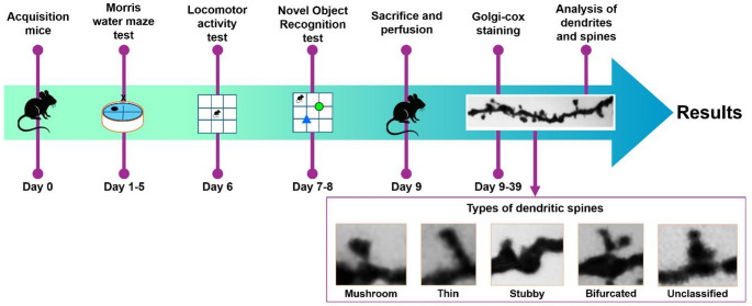
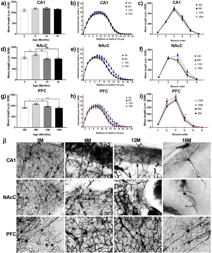
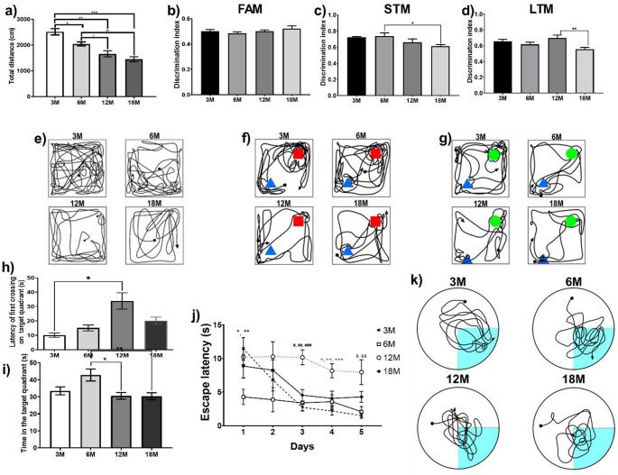
Human populations are experiencing an increase in aging, which is associated with cognitive deficits. Animal models of aging have shown that these behavioral impairments are associated with neuroarchitecture modifications in the prefrontal cortex (PFC) and hippocampus; however, most studies have focused on rats or lack multiple key ages. In this study, we evaluated spatial and recognition memory in male mice at critical ages [3 months (M), 6, 12 and 18] using the Morris water maze (MWM) and novel object recognition test (NORT), respectively. Moreover, we quantified dendritic arborization, spine density and the type of spines in the PFC, CA1 hippocampus and nucleus Accumbens Core (NAcC). Locomotion, assessed in the first phase of NORT, revealed age-dependent reductions. Notably, the 18 M group revealed significant recognition memory deficits. Spatial memory impairments were especially evident at the 12 M group in the MWM. Spine density was increased at 6 M in the NAcC, whereas a reduction was noted at 12 M and 18 M in the PFC. Morphological assessment of spines indicated age-dependent changes, including a notable increase in the proportion of thin spines in the CA1 and PFC regions. However, dendritic arborization remained largely unchanged across the examined brain regions and age groups. Overall, our findings observed age-dependent alterations in memory and morphological alterations in spines in mice, emerging as possible contributors to cognitive decline. These results highlight the potential for anti-aging interventions targeting synaptic structures to enhance cognitive health and extend the healthspan of aging individuals.
期刊介绍:
Brain Structure & Function publishes research that provides insight into brain structure−function relationships. Studies published here integrate data spanning from molecular, cellular, developmental, and systems architecture to the neuroanatomy of behavior and cognitive functions. Manuscripts with focus on the spinal cord or the peripheral nervous system are not accepted for publication. Manuscripts with focus on diseases, animal models of diseases, or disease-related mechanisms are only considered for publication, if the findings provide novel insight into the organization and mechanisms of normal brain structure and function.
 求助内容:
求助内容: 应助结果提醒方式:


