Kejun Li, Yize Han, Yin Zhang, QingMin Ma, Jia Lin Niu, Fang Fan, JianMin Wang
{"title":"2型糖尿病患者角膜及睑板腺形态与糖化血红蛋白水平的相关性分析。","authors":"Kejun Li, Yize Han, Yin Zhang, QingMin Ma, Jia Lin Niu, Fang Fan, JianMin Wang","doi":"10.1186/s12886-025-04273-8","DOIUrl":null,"url":null,"abstract":"<p><strong>Purpose: </strong>To investigate the correlation between corneal nerve and meibomian gland morphological changes and glycated hemoglobin (HbA1c) levels in type 2 diabetes mellitus (T2DM) patients, and to provide an objective basis for early detection of diabetic peripheral neuropathy.</p><p><strong>Methods: </strong>A total of 150 subjects were enrolled, including 130 T2DM patients and 20 healthy controls. T2DM patients were divided into two groups based on HbA1c levels: well-controlled group (HbA1c < 7.0%, n = 65) and poorly-controlled group (HbA1c ≥ 7.0%, n = 65). All subjects underwent comprehensive ophthalmic examinations, including in vivo confocal microscopy (IVCM) for corneal nerve analysis and infrared meibography for meibomian gland evaluation. Disease duration, medication history, and microvascular complications were recorded for all diabetic patients. Age-related meibomian gland changes were controlled by age-matching between groups. Corneal nerve fiber density (CNFD), corneal nerve branch density (CNBD), corneal nerve fiber length (CNFL), and meibomian gland parameters were measured and compared among groups. Correlation analysis was performed between HbA1c levels and ocular surface parameters.</p><p><strong>Results: </strong>Compared with the healthy control group, T2DM patients showed significantly reduced CNFD, CNBD, CNFL, and increased meibomian gland dropout (all P < 0.05). These parameters were significantly worse in the poorly-controlled group compared to the well-controlled group (all P < 0.01). Multiple regression analysis showed that HbA1c levels were independently associated with corneal nerve parameters (CNFD: β = -0.462, P < 0.001; CNBD: β = -0.437, P < 0.001; CNFL: β = -0.443, P < 0.001) and meibomian gland dropout (β = 0.389, P < 0.001) after adjusting for age, disease duration, medication types, and presence of microvascular complications.</p><p><strong>Conclusion: </strong>Corneal nerve fiber and meibomian gland morphology are significantly correlated with HbA1c levels in T2DM patients. These ocular surface changes may serve as early, non-invasive biomarkers for diabetic peripheral neuropathy and could potentially be incorporated into routine diabetic follow-up examinations. Longitudinal studies are needed to establish the causality and evaluate the clinical significance of these findings for individualized ocular surface management in diabetic patients.</p>","PeriodicalId":9058,"journal":{"name":"BMC Ophthalmology","volume":"25 1","pages":"480"},"PeriodicalIF":1.7000,"publicationDate":"2025-08-21","publicationTypes":"Journal Article","fieldsOfStudy":null,"isOpenAccess":false,"openAccessPdf":"https://www.ncbi.nlm.nih.gov/pmc/articles/PMC12369175/pdf/","citationCount":"0","resultStr":"{\"title\":\"Analysis of the correlation between corneal and meibomian gland morphology and glycated hemoglobin levels in type 2 diabetes patients.\",\"authors\":\"Kejun Li, Yize Han, Yin Zhang, QingMin Ma, Jia Lin Niu, Fang Fan, JianMin Wang\",\"doi\":\"10.1186/s12886-025-04273-8\",\"DOIUrl\":null,\"url\":null,\"abstract\":\"<p><strong>Purpose: </strong>To investigate the correlation between corneal nerve and meibomian gland morphological changes and glycated hemoglobin (HbA1c) levels in type 2 diabetes mellitus (T2DM) patients, and to provide an objective basis for early detection of diabetic peripheral neuropathy.</p><p><strong>Methods: </strong>A total of 150 subjects were enrolled, including 130 T2DM patients and 20 healthy controls. T2DM patients were divided into two groups based on HbA1c levels: well-controlled group (HbA1c < 7.0%, n = 65) and poorly-controlled group (HbA1c ≥ 7.0%, n = 65). All subjects underwent comprehensive ophthalmic examinations, including in vivo confocal microscopy (IVCM) for corneal nerve analysis and infrared meibography for meibomian gland evaluation. Disease duration, medication history, and microvascular complications were recorded for all diabetic patients. Age-related meibomian gland changes were controlled by age-matching between groups. Corneal nerve fiber density (CNFD), corneal nerve branch density (CNBD), corneal nerve fiber length (CNFL), and meibomian gland parameters were measured and compared among groups. Correlation analysis was performed between HbA1c levels and ocular surface parameters.</p><p><strong>Results: </strong>Compared with the healthy control group, T2DM patients showed significantly reduced CNFD, CNBD, CNFL, and increased meibomian gland dropout (all P < 0.05). These parameters were significantly worse in the poorly-controlled group compared to the well-controlled group (all P < 0.01). Multiple regression analysis showed that HbA1c levels were independently associated with corneal nerve parameters (CNFD: β = -0.462, P < 0.001; CNBD: β = -0.437, P < 0.001; CNFL: β = -0.443, P < 0.001) and meibomian gland dropout (β = 0.389, P < 0.001) after adjusting for age, disease duration, medication types, and presence of microvascular complications.</p><p><strong>Conclusion: </strong>Corneal nerve fiber and meibomian gland morphology are significantly correlated with HbA1c levels in T2DM patients. These ocular surface changes may serve as early, non-invasive biomarkers for diabetic peripheral neuropathy and could potentially be incorporated into routine diabetic follow-up examinations. Longitudinal studies are needed to establish the causality and evaluate the clinical significance of these findings for individualized ocular surface management in diabetic patients.</p>\",\"PeriodicalId\":9058,\"journal\":{\"name\":\"BMC Ophthalmology\",\"volume\":\"25 1\",\"pages\":\"480\"},\"PeriodicalIF\":1.7000,\"publicationDate\":\"2025-08-21\",\"publicationTypes\":\"Journal Article\",\"fieldsOfStudy\":null,\"isOpenAccess\":false,\"openAccessPdf\":\"https://www.ncbi.nlm.nih.gov/pmc/articles/PMC12369175/pdf/\",\"citationCount\":\"0\",\"resultStr\":null,\"platform\":\"Semanticscholar\",\"paperid\":null,\"PeriodicalName\":\"BMC Ophthalmology\",\"FirstCategoryId\":\"3\",\"ListUrlMain\":\"https://doi.org/10.1186/s12886-025-04273-8\",\"RegionNum\":4,\"RegionCategory\":\"医学\",\"ArticlePicture\":[],\"TitleCN\":null,\"AbstractTextCN\":null,\"PMCID\":null,\"EPubDate\":\"\",\"PubModel\":\"\",\"JCR\":\"Q3\",\"JCRName\":\"OPHTHALMOLOGY\",\"Score\":null,\"Total\":0}","platform":"Semanticscholar","paperid":null,"PeriodicalName":"BMC Ophthalmology","FirstCategoryId":"3","ListUrlMain":"https://doi.org/10.1186/s12886-025-04273-8","RegionNum":4,"RegionCategory":"医学","ArticlePicture":[],"TitleCN":null,"AbstractTextCN":null,"PMCID":null,"EPubDate":"","PubModel":"","JCR":"Q3","JCRName":"OPHTHALMOLOGY","Score":null,"Total":0}
Analysis of the correlation between corneal and meibomian gland morphology and glycated hemoglobin levels in type 2 diabetes patients.
Purpose: To investigate the correlation between corneal nerve and meibomian gland morphological changes and glycated hemoglobin (HbA1c) levels in type 2 diabetes mellitus (T2DM) patients, and to provide an objective basis for early detection of diabetic peripheral neuropathy.
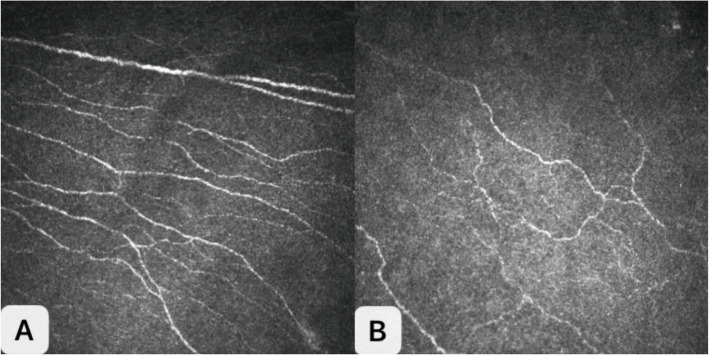
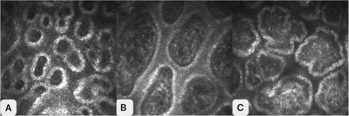
Methods: A total of 150 subjects were enrolled, including 130 T2DM patients and 20 healthy controls. T2DM patients were divided into two groups based on HbA1c levels: well-controlled group (HbA1c < 7.0%, n = 65) and poorly-controlled group (HbA1c ≥ 7.0%, n = 65). All subjects underwent comprehensive ophthalmic examinations, including in vivo confocal microscopy (IVCM) for corneal nerve analysis and infrared meibography for meibomian gland evaluation. Disease duration, medication history, and microvascular complications were recorded for all diabetic patients. Age-related meibomian gland changes were controlled by age-matching between groups. Corneal nerve fiber density (CNFD), corneal nerve branch density (CNBD), corneal nerve fiber length (CNFL), and meibomian gland parameters were measured and compared among groups. Correlation analysis was performed between HbA1c levels and ocular surface parameters.
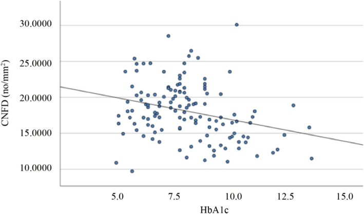
Results: Compared with the healthy control group, T2DM patients showed significantly reduced CNFD, CNBD, CNFL, and increased meibomian gland dropout (all P < 0.05). These parameters were significantly worse in the poorly-controlled group compared to the well-controlled group (all P < 0.01). Multiple regression analysis showed that HbA1c levels were independently associated with corneal nerve parameters (CNFD: β = -0.462, P < 0.001; CNBD: β = -0.437, P < 0.001; CNFL: β = -0.443, P < 0.001) and meibomian gland dropout (β = 0.389, P < 0.001) after adjusting for age, disease duration, medication types, and presence of microvascular complications.
Conclusion: Corneal nerve fiber and meibomian gland morphology are significantly correlated with HbA1c levels in T2DM patients. These ocular surface changes may serve as early, non-invasive biomarkers for diabetic peripheral neuropathy and could potentially be incorporated into routine diabetic follow-up examinations. Longitudinal studies are needed to establish the causality and evaluate the clinical significance of these findings for individualized ocular surface management in diabetic patients.
期刊介绍:
BMC Ophthalmology is an open access, peer-reviewed journal that considers articles on all aspects of the prevention, diagnosis and management of eye disorders, as well as related molecular genetics, pathophysiology, and epidemiology.

 求助内容:
求助内容: 应助结果提醒方式:


