{"title":"孤立检查视觉诱发电位与黄蓝周边镜早期诊断原发性开角型青光眼。","authors":"Qiang Li, Dongyue Liu, Min Zhang","doi":"10.1186/s12886-025-04319-x","DOIUrl":null,"url":null,"abstract":"<p><strong>Background: </strong>Early detection of primary open-angle glaucoma (POAG) is crucial, as current diagnostic methods often miss early-stage damage. Combining blue-on-yellow perimetry with isolated-check visual evoked potential (Ic-VEP) could enhance the sensitivity and specificity of early POAG detection.</p><p><strong>Objective: </strong>To evaluate the diagnostic efficacy of Ic-VEP combined with blue-on-yellow perimetry for early POAG detection.</p><p><strong>Methods: </strong>This study included 66 POAG patients and 35 healthy controls, all of whom underwent comprehensive ophthalmologic assessments, including intraocular pressure (IOP), fundus examination, and optical coherence tomography (OCT) to measure retinal nerve fiber layer (RNFL) and ganglion cell complex (GCC) thickness. Ic-VEP and Humphrey 24 - 2 blue-on-yellow perimetry were performed to assess the sensitivity, specificity, and ROC curve for early POAG diagnosis. The correlation between Ic-VEP. results, GCC thickness, and visual field loss was also analyzed.</p><p><strong>Results: </strong>Ic-VEP demonstrated 74% sensitivity and 91% specificity for detecting POAG, with an area under the ROC curve (AUC) of 0.785, indicating reliable diagnostic performance. The Ic-VEP signal-to-noise ratio (SNR) showed significant correlation with the mean deviation (MD) of blue-on-yellow perimetry and GCC thickness (p < 0.05). The Kappa coefficient for consistency between Ic-VEP and blue-on-yellow perimetry was 0.226 in early-stage POAG, increasing to 0.672 in moderate to severe stages suggesting enhanced diagnostic value in later stages.</p><p><strong>Conclusion: </strong>Combining Ic-VEP with blue-on-yellow perimetry shows promise for enhancing the early diagnosis of POAG, with Ic-VEP's high specificity (91%) complementing the sensitivity of perimetry. This approach could lead to earlier diagnosis and improved patient outcomes.</p>","PeriodicalId":9058,"journal":{"name":"BMC Ophthalmology","volume":"25 1","pages":"483"},"PeriodicalIF":1.7000,"publicationDate":"2025-08-25","publicationTypes":"Journal Article","fieldsOfStudy":null,"isOpenAccess":false,"openAccessPdf":"https://www.ncbi.nlm.nih.gov/pmc/articles/PMC12376748/pdf/","citationCount":"0","resultStr":"{\"title\":\"Early diagnosis of primary open-angle glaucoma using isolated-check visual evoked potential with blue-on-yellow perimetry.\",\"authors\":\"Qiang Li, Dongyue Liu, Min Zhang\",\"doi\":\"10.1186/s12886-025-04319-x\",\"DOIUrl\":null,\"url\":null,\"abstract\":\"<p><strong>Background: </strong>Early detection of primary open-angle glaucoma (POAG) is crucial, as current diagnostic methods often miss early-stage damage. Combining blue-on-yellow perimetry with isolated-check visual evoked potential (Ic-VEP) could enhance the sensitivity and specificity of early POAG detection.</p><p><strong>Objective: </strong>To evaluate the diagnostic efficacy of Ic-VEP combined with blue-on-yellow perimetry for early POAG detection.</p><p><strong>Methods: </strong>This study included 66 POAG patients and 35 healthy controls, all of whom underwent comprehensive ophthalmologic assessments, including intraocular pressure (IOP), fundus examination, and optical coherence tomography (OCT) to measure retinal nerve fiber layer (RNFL) and ganglion cell complex (GCC) thickness. Ic-VEP and Humphrey 24 - 2 blue-on-yellow perimetry were performed to assess the sensitivity, specificity, and ROC curve for early POAG diagnosis. The correlation between Ic-VEP. results, GCC thickness, and visual field loss was also analyzed.</p><p><strong>Results: </strong>Ic-VEP demonstrated 74% sensitivity and 91% specificity for detecting POAG, with an area under the ROC curve (AUC) of 0.785, indicating reliable diagnostic performance. The Ic-VEP signal-to-noise ratio (SNR) showed significant correlation with the mean deviation (MD) of blue-on-yellow perimetry and GCC thickness (p < 0.05). The Kappa coefficient for consistency between Ic-VEP and blue-on-yellow perimetry was 0.226 in early-stage POAG, increasing to 0.672 in moderate to severe stages suggesting enhanced diagnostic value in later stages.</p><p><strong>Conclusion: </strong>Combining Ic-VEP with blue-on-yellow perimetry shows promise for enhancing the early diagnosis of POAG, with Ic-VEP's high specificity (91%) complementing the sensitivity of perimetry. This approach could lead to earlier diagnosis and improved patient outcomes.</p>\",\"PeriodicalId\":9058,\"journal\":{\"name\":\"BMC Ophthalmology\",\"volume\":\"25 1\",\"pages\":\"483\"},\"PeriodicalIF\":1.7000,\"publicationDate\":\"2025-08-25\",\"publicationTypes\":\"Journal Article\",\"fieldsOfStudy\":null,\"isOpenAccess\":false,\"openAccessPdf\":\"https://www.ncbi.nlm.nih.gov/pmc/articles/PMC12376748/pdf/\",\"citationCount\":\"0\",\"resultStr\":null,\"platform\":\"Semanticscholar\",\"paperid\":null,\"PeriodicalName\":\"BMC Ophthalmology\",\"FirstCategoryId\":\"3\",\"ListUrlMain\":\"https://doi.org/10.1186/s12886-025-04319-x\",\"RegionNum\":4,\"RegionCategory\":\"医学\",\"ArticlePicture\":[],\"TitleCN\":null,\"AbstractTextCN\":null,\"PMCID\":null,\"EPubDate\":\"\",\"PubModel\":\"\",\"JCR\":\"Q3\",\"JCRName\":\"OPHTHALMOLOGY\",\"Score\":null,\"Total\":0}","platform":"Semanticscholar","paperid":null,"PeriodicalName":"BMC Ophthalmology","FirstCategoryId":"3","ListUrlMain":"https://doi.org/10.1186/s12886-025-04319-x","RegionNum":4,"RegionCategory":"医学","ArticlePicture":[],"TitleCN":null,"AbstractTextCN":null,"PMCID":null,"EPubDate":"","PubModel":"","JCR":"Q3","JCRName":"OPHTHALMOLOGY","Score":null,"Total":0}
Early diagnosis of primary open-angle glaucoma using isolated-check visual evoked potential with blue-on-yellow perimetry.
Background: Early detection of primary open-angle glaucoma (POAG) is crucial, as current diagnostic methods often miss early-stage damage. Combining blue-on-yellow perimetry with isolated-check visual evoked potential (Ic-VEP) could enhance the sensitivity and specificity of early POAG detection.
Objective: To evaluate the diagnostic efficacy of Ic-VEP combined with blue-on-yellow perimetry for early POAG detection.
Methods: This study included 66 POAG patients and 35 healthy controls, all of whom underwent comprehensive ophthalmologic assessments, including intraocular pressure (IOP), fundus examination, and optical coherence tomography (OCT) to measure retinal nerve fiber layer (RNFL) and ganglion cell complex (GCC) thickness. Ic-VEP and Humphrey 24 - 2 blue-on-yellow perimetry were performed to assess the sensitivity, specificity, and ROC curve for early POAG diagnosis. The correlation between Ic-VEP. results, GCC thickness, and visual field loss was also analyzed.
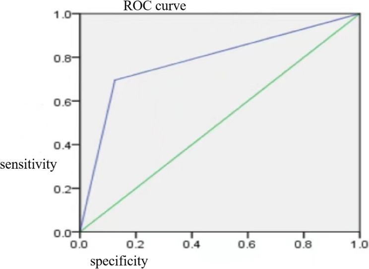
Results: Ic-VEP demonstrated 74% sensitivity and 91% specificity for detecting POAG, with an area under the ROC curve (AUC) of 0.785, indicating reliable diagnostic performance. The Ic-VEP signal-to-noise ratio (SNR) showed significant correlation with the mean deviation (MD) of blue-on-yellow perimetry and GCC thickness (p < 0.05). The Kappa coefficient for consistency between Ic-VEP and blue-on-yellow perimetry was 0.226 in early-stage POAG, increasing to 0.672 in moderate to severe stages suggesting enhanced diagnostic value in later stages.
Conclusion: Combining Ic-VEP with blue-on-yellow perimetry shows promise for enhancing the early diagnosis of POAG, with Ic-VEP's high specificity (91%) complementing the sensitivity of perimetry. This approach could lead to earlier diagnosis and improved patient outcomes.
期刊介绍:
BMC Ophthalmology is an open access, peer-reviewed journal that considers articles on all aspects of the prevention, diagnosis and management of eye disorders, as well as related molecular genetics, pathophysiology, and epidemiology.

 求助内容:
求助内容: 应助结果提醒方式:


