{"title":"应用倒置限制性膜瓣技术观察特发性黄斑裂孔手术后神经节细胞层的变化。","authors":"Mehmet Önen, Muzaffer Şahin, Gökhan Çelik","doi":"10.1186/s12886-025-04317-z","DOIUrl":null,"url":null,"abstract":"<p><strong>Purpose: </strong>The purpose of this study is to compare the ganglion cell layer changes following temporal inverted internal limiting membrane flap (i-ILMF) surgery for idiopathic macular hole (IMH).</p><p><strong>Methods: </strong>This retrospective study included 50 eyes that underwent vitrectomy with a 2.5-disc-diameter temporal inverted internal limiting membrane flap (i-ILMF) technique. Demographic, functional, and anatomical data were collected before and after the surgery. The best corrected visual acuity (BCVA) and optical coherence tomography (OCT) findings such as ganglion cell layer -inner plexiform layer (GCL-IPL) thickness and hole related parameters/indexes were compared in the preoperative period and 6th month after surgery.</p><p><strong>Results: </strong>The average age of the patients was 68.8 ± 10.31 years, and the average duration of visual loss was 10.95 ± 6.54 months. The average GCL-IPL thickness increased significantly from 57.98 ± 21.43 μm to 68.74 ± 13.62 μm at 6 months after surgery (p < 0.001). The nasal GCL-IPL thickness was significantly increased from 56.94 ± 24.18 μm to 73.10 ± 15.39 μm after 6 months after surgery (p < 0.001).</p><p><strong>Conclusion: </strong>The temporal i-ILMF technique not only leads to high anatomical success and visual improvement but also results in a significant increase in GCL-IPL thickness postoperatively, suggesting a unique structural response to this method.</p>","PeriodicalId":9058,"journal":{"name":"BMC Ophthalmology","volume":"25 1","pages":"481"},"PeriodicalIF":1.7000,"publicationDate":"2025-08-21","publicationTypes":"Journal Article","fieldsOfStudy":null,"isOpenAccess":false,"openAccessPdf":"https://www.ncbi.nlm.nih.gov/pmc/articles/PMC12372387/pdf/","citationCount":"0","resultStr":"{\"title\":\"Ganglion cell layer changes following the idiopathic macular hole surgery using inverted limiting membrane flap technique.\",\"authors\":\"Mehmet Önen, Muzaffer Şahin, Gökhan Çelik\",\"doi\":\"10.1186/s12886-025-04317-z\",\"DOIUrl\":null,\"url\":null,\"abstract\":\"<p><strong>Purpose: </strong>The purpose of this study is to compare the ganglion cell layer changes following temporal inverted internal limiting membrane flap (i-ILMF) surgery for idiopathic macular hole (IMH).</p><p><strong>Methods: </strong>This retrospective study included 50 eyes that underwent vitrectomy with a 2.5-disc-diameter temporal inverted internal limiting membrane flap (i-ILMF) technique. Demographic, functional, and anatomical data were collected before and after the surgery. The best corrected visual acuity (BCVA) and optical coherence tomography (OCT) findings such as ganglion cell layer -inner plexiform layer (GCL-IPL) thickness and hole related parameters/indexes were compared in the preoperative period and 6th month after surgery.</p><p><strong>Results: </strong>The average age of the patients was 68.8 ± 10.31 years, and the average duration of visual loss was 10.95 ± 6.54 months. The average GCL-IPL thickness increased significantly from 57.98 ± 21.43 μm to 68.74 ± 13.62 μm at 6 months after surgery (p < 0.001). The nasal GCL-IPL thickness was significantly increased from 56.94 ± 24.18 μm to 73.10 ± 15.39 μm after 6 months after surgery (p < 0.001).</p><p><strong>Conclusion: </strong>The temporal i-ILMF technique not only leads to high anatomical success and visual improvement but also results in a significant increase in GCL-IPL thickness postoperatively, suggesting a unique structural response to this method.</p>\",\"PeriodicalId\":9058,\"journal\":{\"name\":\"BMC Ophthalmology\",\"volume\":\"25 1\",\"pages\":\"481\"},\"PeriodicalIF\":1.7000,\"publicationDate\":\"2025-08-21\",\"publicationTypes\":\"Journal Article\",\"fieldsOfStudy\":null,\"isOpenAccess\":false,\"openAccessPdf\":\"https://www.ncbi.nlm.nih.gov/pmc/articles/PMC12372387/pdf/\",\"citationCount\":\"0\",\"resultStr\":null,\"platform\":\"Semanticscholar\",\"paperid\":null,\"PeriodicalName\":\"BMC Ophthalmology\",\"FirstCategoryId\":\"3\",\"ListUrlMain\":\"https://doi.org/10.1186/s12886-025-04317-z\",\"RegionNum\":4,\"RegionCategory\":\"医学\",\"ArticlePicture\":[],\"TitleCN\":null,\"AbstractTextCN\":null,\"PMCID\":null,\"EPubDate\":\"\",\"PubModel\":\"\",\"JCR\":\"Q3\",\"JCRName\":\"OPHTHALMOLOGY\",\"Score\":null,\"Total\":0}","platform":"Semanticscholar","paperid":null,"PeriodicalName":"BMC Ophthalmology","FirstCategoryId":"3","ListUrlMain":"https://doi.org/10.1186/s12886-025-04317-z","RegionNum":4,"RegionCategory":"医学","ArticlePicture":[],"TitleCN":null,"AbstractTextCN":null,"PMCID":null,"EPubDate":"","PubModel":"","JCR":"Q3","JCRName":"OPHTHALMOLOGY","Score":null,"Total":0}
Ganglion cell layer changes following the idiopathic macular hole surgery using inverted limiting membrane flap technique.
Purpose: The purpose of this study is to compare the ganglion cell layer changes following temporal inverted internal limiting membrane flap (i-ILMF) surgery for idiopathic macular hole (IMH).
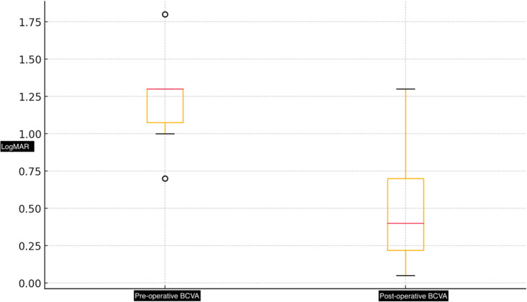
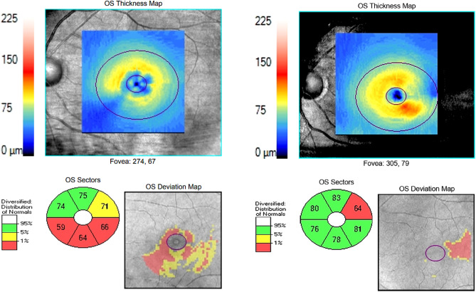
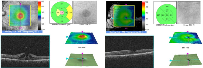
Methods: This retrospective study included 50 eyes that underwent vitrectomy with a 2.5-disc-diameter temporal inverted internal limiting membrane flap (i-ILMF) technique. Demographic, functional, and anatomical data were collected before and after the surgery. The best corrected visual acuity (BCVA) and optical coherence tomography (OCT) findings such as ganglion cell layer -inner plexiform layer (GCL-IPL) thickness and hole related parameters/indexes were compared in the preoperative period and 6th month after surgery.
Results: The average age of the patients was 68.8 ± 10.31 years, and the average duration of visual loss was 10.95 ± 6.54 months. The average GCL-IPL thickness increased significantly from 57.98 ± 21.43 μm to 68.74 ± 13.62 μm at 6 months after surgery (p < 0.001). The nasal GCL-IPL thickness was significantly increased from 56.94 ± 24.18 μm to 73.10 ± 15.39 μm after 6 months after surgery (p < 0.001).
Conclusion: The temporal i-ILMF technique not only leads to high anatomical success and visual improvement but also results in a significant increase in GCL-IPL thickness postoperatively, suggesting a unique structural response to this method.
期刊介绍:
BMC Ophthalmology is an open access, peer-reviewed journal that considers articles on all aspects of the prevention, diagnosis and management of eye disorders, as well as related molecular genetics, pathophysiology, and epidemiology.

 求助内容:
求助内容: 应助结果提醒方式:


