Shuai Zhang, Na Chang, Xinxin Yu, Bing Kang, Ru Tan
{"title":"CT血管造影血管周围脂肪密度与腹主动脉瘤进展的关系。","authors":"Shuai Zhang, Na Chang, Xinxin Yu, Bing Kang, Ru Tan","doi":"10.1186/s12880-025-01895-8","DOIUrl":null,"url":null,"abstract":"<p><strong>Background: </strong>Perivascular adipose tissue has been shown to play a role in cardiovascular disease. This provides evidences that perivascular fat density (PFD) may have a correlation with abdominal aortic aneurysm (AAA). The aim of study was to investigate the association between PFD on computed tomography angiography (CTA) and AAA expanding rate.</p><p><strong>Methods: </strong>A total of 144 patients with AAA who underwent at least two computed tomography angiography (CTA) examinations at intervals of ≥ 6 months between January 2014 and June 2023 were included. The patients were divided into slowly and rapidly expanding AAA groups according to the median value of AAA expansion rate. The clinical and CTA characteristics of both groups were compared. The relationships between AAA diameter, AAA volume, expansion rate, and PFD were tested using the pearson coefficient.</p><p><strong>Results: </strong>Compared with the slowly expanding group, patients with rapidly expanding AAA had a significantly higher prevalence of hypertension (77.8% versus 55.6%; P = 0.005), a significantly lower prevalence of diabetes (26.4% versus 47.2%; P < 0.010), and a higher PFD at baseline (-72.6 ± 9.7 HU vs. -81.2 ± 7.9 HU; P < 0.001). In the whole group, slowly expanding group, and rapidly expanding group, PFD at baseline was positively correlated with AAA diameter at baseline (P < 0.001), AAA volume at baseline (P < 0.001), and expansion rate (P < 0.001). A positive correlation was observed between increasing PFD and expansion rate (P < 0.05).</p><p><strong>Conclusions: </strong>A higher PFD on CTA was found to be related to a rapidly expanding AAA. Thus, PFD may become a non-invasive and potential image marker for predicting and treating AAA progression.</p><p><strong>Clinical trial number: </strong>Not applicable. This research is a retrospective analysis.</p>","PeriodicalId":9020,"journal":{"name":"BMC Medical Imaging","volume":"25 1","pages":"357"},"PeriodicalIF":3.2000,"publicationDate":"2025-08-29","publicationTypes":"Journal Article","fieldsOfStudy":null,"isOpenAccess":false,"openAccessPdf":"https://www.ncbi.nlm.nih.gov/pmc/articles/PMC12395999/pdf/","citationCount":"0","resultStr":"{\"title\":\"Association between perivascular fat density on CT angiography and abdominal aortic aneurysm progression.\",\"authors\":\"Shuai Zhang, Na Chang, Xinxin Yu, Bing Kang, Ru Tan\",\"doi\":\"10.1186/s12880-025-01895-8\",\"DOIUrl\":null,\"url\":null,\"abstract\":\"<p><strong>Background: </strong>Perivascular adipose tissue has been shown to play a role in cardiovascular disease. This provides evidences that perivascular fat density (PFD) may have a correlation with abdominal aortic aneurysm (AAA). The aim of study was to investigate the association between PFD on computed tomography angiography (CTA) and AAA expanding rate.</p><p><strong>Methods: </strong>A total of 144 patients with AAA who underwent at least two computed tomography angiography (CTA) examinations at intervals of ≥ 6 months between January 2014 and June 2023 were included. The patients were divided into slowly and rapidly expanding AAA groups according to the median value of AAA expansion rate. The clinical and CTA characteristics of both groups were compared. The relationships between AAA diameter, AAA volume, expansion rate, and PFD were tested using the pearson coefficient.</p><p><strong>Results: </strong>Compared with the slowly expanding group, patients with rapidly expanding AAA had a significantly higher prevalence of hypertension (77.8% versus 55.6%; P = 0.005), a significantly lower prevalence of diabetes (26.4% versus 47.2%; P < 0.010), and a higher PFD at baseline (-72.6 ± 9.7 HU vs. -81.2 ± 7.9 HU; P < 0.001). In the whole group, slowly expanding group, and rapidly expanding group, PFD at baseline was positively correlated with AAA diameter at baseline (P < 0.001), AAA volume at baseline (P < 0.001), and expansion rate (P < 0.001). A positive correlation was observed between increasing PFD and expansion rate (P < 0.05).</p><p><strong>Conclusions: </strong>A higher PFD on CTA was found to be related to a rapidly expanding AAA. Thus, PFD may become a non-invasive and potential image marker for predicting and treating AAA progression.</p><p><strong>Clinical trial number: </strong>Not applicable. This research is a retrospective analysis.</p>\",\"PeriodicalId\":9020,\"journal\":{\"name\":\"BMC Medical Imaging\",\"volume\":\"25 1\",\"pages\":\"357\"},\"PeriodicalIF\":3.2000,\"publicationDate\":\"2025-08-29\",\"publicationTypes\":\"Journal Article\",\"fieldsOfStudy\":null,\"isOpenAccess\":false,\"openAccessPdf\":\"https://www.ncbi.nlm.nih.gov/pmc/articles/PMC12395999/pdf/\",\"citationCount\":\"0\",\"resultStr\":null,\"platform\":\"Semanticscholar\",\"paperid\":null,\"PeriodicalName\":\"BMC Medical Imaging\",\"FirstCategoryId\":\"3\",\"ListUrlMain\":\"https://doi.org/10.1186/s12880-025-01895-8\",\"RegionNum\":3,\"RegionCategory\":\"医学\",\"ArticlePicture\":[],\"TitleCN\":null,\"AbstractTextCN\":null,\"PMCID\":null,\"EPubDate\":\"\",\"PubModel\":\"\",\"JCR\":\"Q2\",\"JCRName\":\"RADIOLOGY, NUCLEAR MEDICINE & MEDICAL IMAGING\",\"Score\":null,\"Total\":0}","platform":"Semanticscholar","paperid":null,"PeriodicalName":"BMC Medical Imaging","FirstCategoryId":"3","ListUrlMain":"https://doi.org/10.1186/s12880-025-01895-8","RegionNum":3,"RegionCategory":"医学","ArticlePicture":[],"TitleCN":null,"AbstractTextCN":null,"PMCID":null,"EPubDate":"","PubModel":"","JCR":"Q2","JCRName":"RADIOLOGY, NUCLEAR MEDICINE & MEDICAL IMAGING","Score":null,"Total":0}
Association between perivascular fat density on CT angiography and abdominal aortic aneurysm progression.
Background: Perivascular adipose tissue has been shown to play a role in cardiovascular disease. This provides evidences that perivascular fat density (PFD) may have a correlation with abdominal aortic aneurysm (AAA). The aim of study was to investigate the association between PFD on computed tomography angiography (CTA) and AAA expanding rate.
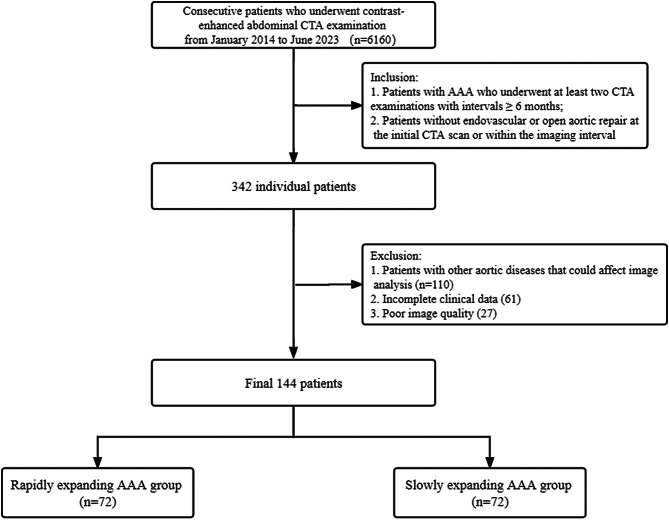
Methods: A total of 144 patients with AAA who underwent at least two computed tomography angiography (CTA) examinations at intervals of ≥ 6 months between January 2014 and June 2023 were included. The patients were divided into slowly and rapidly expanding AAA groups according to the median value of AAA expansion rate. The clinical and CTA characteristics of both groups were compared. The relationships between AAA diameter, AAA volume, expansion rate, and PFD were tested using the pearson coefficient.
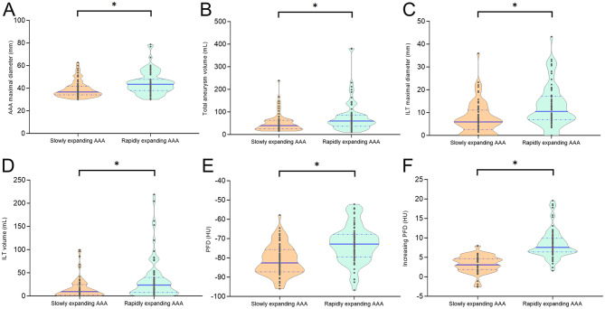
Results: Compared with the slowly expanding group, patients with rapidly expanding AAA had a significantly higher prevalence of hypertension (77.8% versus 55.6%; P = 0.005), a significantly lower prevalence of diabetes (26.4% versus 47.2%; P < 0.010), and a higher PFD at baseline (-72.6 ± 9.7 HU vs. -81.2 ± 7.9 HU; P < 0.001). In the whole group, slowly expanding group, and rapidly expanding group, PFD at baseline was positively correlated with AAA diameter at baseline (P < 0.001), AAA volume at baseline (P < 0.001), and expansion rate (P < 0.001). A positive correlation was observed between increasing PFD and expansion rate (P < 0.05).
Conclusions: A higher PFD on CTA was found to be related to a rapidly expanding AAA. Thus, PFD may become a non-invasive and potential image marker for predicting and treating AAA progression.
Clinical trial number: Not applicable. This research is a retrospective analysis.
期刊介绍:
BMC Medical Imaging is an open access journal publishing original peer-reviewed research articles in the development, evaluation, and use of imaging techniques and image processing tools to diagnose and manage disease.

 求助内容:
求助内容: 应助结果提醒方式:


