{"title":"多参数MRI对原发性中枢神经系统淋巴瘤和非典型胶质母细胞瘤的鉴别诊断:结合DWI、DCE-MRI和造影剂预负荷DSC-PWI的分析","authors":"Lan Yu, Shujie Yu, Feng Wang, Xiaofang Zhou, Feiman Yang, Dairong Cao, Zhen Xing","doi":"10.1186/s12880-025-01886-9","DOIUrl":null,"url":null,"abstract":"<p><strong>Purpose: </strong>The differential diagnosis of primary central nervous system lymphoma (PCNSL) and atypical glioblastoma (aGBM) exhibiting homogeneous enhancement and negligible necrosis poses a significant challenge for conventional MRI. The study aims to investigate diffusion-weighted imaging (DWI), dynamic contrast-enhanced (DCE) MRI, and contrast agent (CA) preload dynamic susceptibility contrast perfusion-weighted imaging (DSC-PWI) to differentiate aGBM and PCNSL.</p><p><strong>Materials and methods: </strong>This retrospective study analyzed 27 patients with aGBM (solid enhancement without visible necrosis) and 105 patients with PCNSL, all undergoing preoperative DWI, DCE-MRI, and CA preload DSC-PWI. The relative apparent diffusion coefficient (rADC) and relative cerebral blood volume (rCBV) were obtained from DWI and DSC-PWI. The pharmacokinetic parameters (Ktrans, Ve, Kep, and iAUC) were acquired using DCE-MRI. The independent-samples t-test and Mann-Whitney U test were utilized to compare parameters. A binary logistic regression analysis was performed to assess the combined effect of various parameters. Before regression analysis, collinearity analysis of parameters was performed. The diagnostic capability of each parameter and their combination were evaluated by receiver operating characteristic (ROC) with area under the curve (AUC) and compared with DeLong test.</p><p><strong>Results: </strong>In comparison to aGBM, the Ktrans, Ve, and iAUC were significantly elevated in PCNSL, whereas the rCBV and rADC were significantly lower (p < 0.05 for all comparisons). Meanwhile, these parameters allowed excellent diagnostic performance (AUC = 0.817 [rCBV], 0.751 [rADC], 0.808 [Ktrans], 0.765 [Ve], and 0.801 [iAUC]; DeLong test, p > 0.05 for all comparisons). Notably, the combination of all these parameters significantly increased the probability of distinguishing aGBM from PCNSL (AUC = 0.966).</p><p><strong>Conclusions: </strong>DWI, DCE-MRI, and CA preload DSC-PWI can effectively differentiate aGBM from PCNSL, and the combination of all three techniques significantly enhances the discriminatory efficacy.</p>","PeriodicalId":9020,"journal":{"name":"BMC Medical Imaging","volume":"25 1","pages":"345"},"PeriodicalIF":3.2000,"publicationDate":"2025-08-25","publicationTypes":"Journal Article","fieldsOfStudy":null,"isOpenAccess":false,"openAccessPdf":"https://www.ncbi.nlm.nih.gov/pmc/articles/PMC12376417/pdf/","citationCount":"0","resultStr":"{\"title\":\"Multiparametric MRI for differential diagnosis of primary central nervous system lymphoma and atypical glioblastoma: an analysis incorporating DWI, DCE-MRI, and contrast agent preload DSC-PWI.\",\"authors\":\"Lan Yu, Shujie Yu, Feng Wang, Xiaofang Zhou, Feiman Yang, Dairong Cao, Zhen Xing\",\"doi\":\"10.1186/s12880-025-01886-9\",\"DOIUrl\":null,\"url\":null,\"abstract\":\"<p><strong>Purpose: </strong>The differential diagnosis of primary central nervous system lymphoma (PCNSL) and atypical glioblastoma (aGBM) exhibiting homogeneous enhancement and negligible necrosis poses a significant challenge for conventional MRI. The study aims to investigate diffusion-weighted imaging (DWI), dynamic contrast-enhanced (DCE) MRI, and contrast agent (CA) preload dynamic susceptibility contrast perfusion-weighted imaging (DSC-PWI) to differentiate aGBM and PCNSL.</p><p><strong>Materials and methods: </strong>This retrospective study analyzed 27 patients with aGBM (solid enhancement without visible necrosis) and 105 patients with PCNSL, all undergoing preoperative DWI, DCE-MRI, and CA preload DSC-PWI. The relative apparent diffusion coefficient (rADC) and relative cerebral blood volume (rCBV) were obtained from DWI and DSC-PWI. The pharmacokinetic parameters (Ktrans, Ve, Kep, and iAUC) were acquired using DCE-MRI. The independent-samples t-test and Mann-Whitney U test were utilized to compare parameters. A binary logistic regression analysis was performed to assess the combined effect of various parameters. Before regression analysis, collinearity analysis of parameters was performed. The diagnostic capability of each parameter and their combination were evaluated by receiver operating characteristic (ROC) with area under the curve (AUC) and compared with DeLong test.</p><p><strong>Results: </strong>In comparison to aGBM, the Ktrans, Ve, and iAUC were significantly elevated in PCNSL, whereas the rCBV and rADC were significantly lower (p < 0.05 for all comparisons). Meanwhile, these parameters allowed excellent diagnostic performance (AUC = 0.817 [rCBV], 0.751 [rADC], 0.808 [Ktrans], 0.765 [Ve], and 0.801 [iAUC]; DeLong test, p > 0.05 for all comparisons). Notably, the combination of all these parameters significantly increased the probability of distinguishing aGBM from PCNSL (AUC = 0.966).</p><p><strong>Conclusions: </strong>DWI, DCE-MRI, and CA preload DSC-PWI can effectively differentiate aGBM from PCNSL, and the combination of all three techniques significantly enhances the discriminatory efficacy.</p>\",\"PeriodicalId\":9020,\"journal\":{\"name\":\"BMC Medical Imaging\",\"volume\":\"25 1\",\"pages\":\"345\"},\"PeriodicalIF\":3.2000,\"publicationDate\":\"2025-08-25\",\"publicationTypes\":\"Journal Article\",\"fieldsOfStudy\":null,\"isOpenAccess\":false,\"openAccessPdf\":\"https://www.ncbi.nlm.nih.gov/pmc/articles/PMC12376417/pdf/\",\"citationCount\":\"0\",\"resultStr\":null,\"platform\":\"Semanticscholar\",\"paperid\":null,\"PeriodicalName\":\"BMC Medical Imaging\",\"FirstCategoryId\":\"3\",\"ListUrlMain\":\"https://doi.org/10.1186/s12880-025-01886-9\",\"RegionNum\":3,\"RegionCategory\":\"医学\",\"ArticlePicture\":[],\"TitleCN\":null,\"AbstractTextCN\":null,\"PMCID\":null,\"EPubDate\":\"\",\"PubModel\":\"\",\"JCR\":\"Q2\",\"JCRName\":\"RADIOLOGY, NUCLEAR MEDICINE & MEDICAL IMAGING\",\"Score\":null,\"Total\":0}","platform":"Semanticscholar","paperid":null,"PeriodicalName":"BMC Medical Imaging","FirstCategoryId":"3","ListUrlMain":"https://doi.org/10.1186/s12880-025-01886-9","RegionNum":3,"RegionCategory":"医学","ArticlePicture":[],"TitleCN":null,"AbstractTextCN":null,"PMCID":null,"EPubDate":"","PubModel":"","JCR":"Q2","JCRName":"RADIOLOGY, NUCLEAR MEDICINE & MEDICAL IMAGING","Score":null,"Total":0}
引用次数: 0
摘要
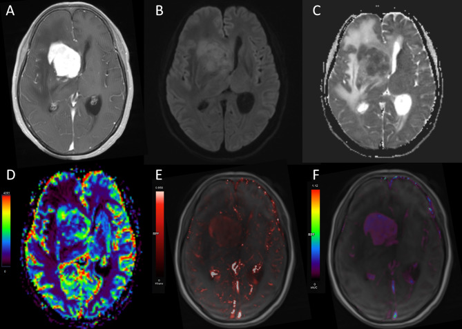
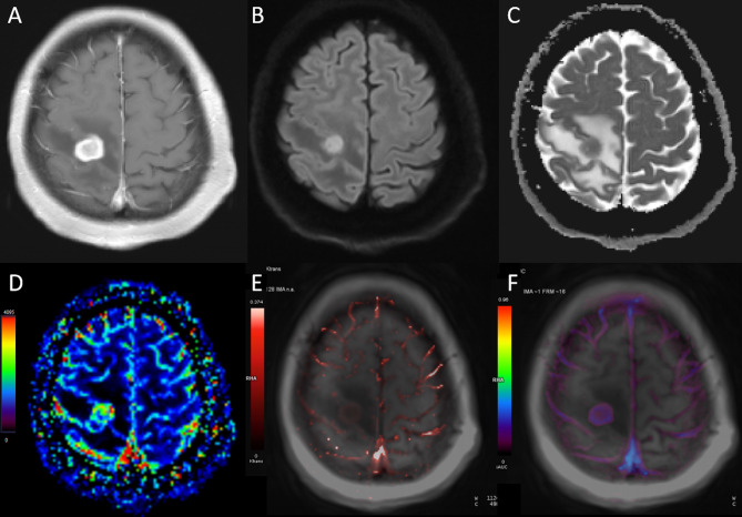
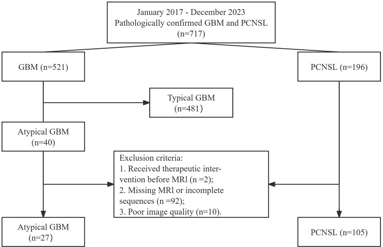
目的:原发性中枢神经系统淋巴瘤(PCNSL)和非典型胶质母细胞瘤(aGBM)表现为均匀强化和可忽略的坏死,其鉴别诊断对传统MRI提出了重大挑战。本研究旨在探讨扩散加权成像(DWI)、动态对比增强(DCE) MRI和造影剂(CA)预负荷动态敏感性对比灌注加权成像(DSC-PWI)对aGBM和PCNSL的鉴别价值。材料和方法:本回顾性研究分析了27例aGBM(实性强化无可见坏死)和105例PCNSL患者,所有患者术前均进行了DWI、DCE-MRI和CA预载DSC-PWI检查。DWI和DSC-PWI分别获取相对表观扩散系数(rADC)和相对脑血容量(rCBV)。通过DCE-MRI获得药代动力学参数(Ktrans, Ve, Kep, iAUC)。采用独立样本t检验和Mann-Whitney U检验进行参数比较。采用二元logistic回归分析评估各参数的综合影响。在进行回归分析之前,对参数进行共线性分析。采用受试者工作特征(ROC)和曲线下面积(AUC)评价各参数及其组合的诊断能力,并与DeLong试验进行比较。结果:与aGBM相比,PCNSL患者Ktrans、Ve、iAUC均显著升高,rCBV、rADC均显著降低(p < 0.05)。值得注意的是,所有这些参数的组合显著提高了aGBM与PCNSL的区分概率(AUC = 0.966)。结论:DWI、DCE-MRI、CA预载DSC-PWI可有效鉴别aGBM与PCNSL,三者联合应用可显著增强鉴别效果。
Multiparametric MRI for differential diagnosis of primary central nervous system lymphoma and atypical glioblastoma: an analysis incorporating DWI, DCE-MRI, and contrast agent preload DSC-PWI.
Purpose: The differential diagnosis of primary central nervous system lymphoma (PCNSL) and atypical glioblastoma (aGBM) exhibiting homogeneous enhancement and negligible necrosis poses a significant challenge for conventional MRI. The study aims to investigate diffusion-weighted imaging (DWI), dynamic contrast-enhanced (DCE) MRI, and contrast agent (CA) preload dynamic susceptibility contrast perfusion-weighted imaging (DSC-PWI) to differentiate aGBM and PCNSL.
Materials and methods: This retrospective study analyzed 27 patients with aGBM (solid enhancement without visible necrosis) and 105 patients with PCNSL, all undergoing preoperative DWI, DCE-MRI, and CA preload DSC-PWI. The relative apparent diffusion coefficient (rADC) and relative cerebral blood volume (rCBV) were obtained from DWI and DSC-PWI. The pharmacokinetic parameters (Ktrans, Ve, Kep, and iAUC) were acquired using DCE-MRI. The independent-samples t-test and Mann-Whitney U test were utilized to compare parameters. A binary logistic regression analysis was performed to assess the combined effect of various parameters. Before regression analysis, collinearity analysis of parameters was performed. The diagnostic capability of each parameter and their combination were evaluated by receiver operating characteristic (ROC) with area under the curve (AUC) and compared with DeLong test.
Results: In comparison to aGBM, the Ktrans, Ve, and iAUC were significantly elevated in PCNSL, whereas the rCBV and rADC were significantly lower (p < 0.05 for all comparisons). Meanwhile, these parameters allowed excellent diagnostic performance (AUC = 0.817 [rCBV], 0.751 [rADC], 0.808 [Ktrans], 0.765 [Ve], and 0.801 [iAUC]; DeLong test, p > 0.05 for all comparisons). Notably, the combination of all these parameters significantly increased the probability of distinguishing aGBM from PCNSL (AUC = 0.966).
Conclusions: DWI, DCE-MRI, and CA preload DSC-PWI can effectively differentiate aGBM from PCNSL, and the combination of all three techniques significantly enhances the discriminatory efficacy.
期刊介绍:
BMC Medical Imaging is an open access journal publishing original peer-reviewed research articles in the development, evaluation, and use of imaging techniques and image processing tools to diagnose and manage disease.

 求助内容:
求助内容: 应助结果提醒方式:


