Peng An, Nan Jiang, Jinyan Li, Wei Li, Kun Zhou, Jiaxiang Xin, Peihang Jing, Lixin Sun
{"title":"扩散加权成像和动态增强MRI对腺样囊性癌病理分级的诊断价值。","authors":"Peng An, Nan Jiang, Jinyan Li, Wei Li, Kun Zhou, Jiaxiang Xin, Peihang Jing, Lixin Sun","doi":"10.1186/s12880-025-01898-5","DOIUrl":null,"url":null,"abstract":"<p><strong>Objectives: </strong>The combination of dynamic-contrast-enhanced-magnetic-resonance-imaging (DCE-MRI) with the apparent-diffusion-coefficient (ADC) holds significant value for predicting tumor pathological outcomes. This study pioneers the combined application of DCE-MRI and ADC parameters to evaluate their utility in differentiating histopathological grades of adenoid cystic carcinoma (ACC).</p><p><strong>Methods: </strong>Retrospective diagnosis of 23 ear and temporal ACC patients was confirmed based on surgical pathology from March 2020 to April 2024. All patients underwent routine MRI, DWI, and DCE-MRI scans within one week before surgery. The lesion ADC values and DCE-MRI perfusion parameters, including Ve, Kep, Ktrans, and iAUC, were measured. Consistency tests were conducted on the measurements done by two physicians. The ADC values and DCE-MRI perfusion parameters between different pathological grades were compared. The correlation among all parameters and ACC pathological grading were analyzed, and the diagnostic accuracy of each parameter was assessed using receiver-operating-characteristic (ROC) curves.</p><p><strong>Results: </strong>The measurements from the two observers showed high consistency (ICC > 0.9). Ktrans, iAUC, and ADC values demonstrated significant differences between different pathological grades (P < .01, P < .05, P < .05). Correlation analysis indicated that Ktrans, Kep, Ve, and iAUC were positively correlated with ACC pathological grading, and Ktrans demonstrated the most robust correlation (correlation coefficient r = .578, P < .01). In contrast, ADC values were markedly and negatively correlated with pathological grading (r=-.470, P < .05). In ROC analysis, the area-under-the-curve (AUC) for Ktrans, iAUC, and ADC were 0.841, 0.790, and 0.778, respectively, all higher than those for Kep and Ve. Ktrans showed the best diagnostic performance.</p><p><strong>Conclusion: </strong>Combining DCE-MRI perfusion parameters with ADC values provides a non-invasive and effective method for preoperative grading of ACC, with Ktrans, iAUC, and ADC showing strong diagnostic potential. These findings support more accurate tumor characterization and personalized treatment planning, warranting further validation in larger prospective studies.</p><p><strong>Clinical trial number: </strong>Not applicable.</p>","PeriodicalId":9020,"journal":{"name":"BMC Medical Imaging","volume":"25 1","pages":"359"},"PeriodicalIF":3.2000,"publicationDate":"2025-08-30","publicationTypes":"Journal Article","fieldsOfStudy":null,"isOpenAccess":false,"openAccessPdf":"https://www.ncbi.nlm.nih.gov/pmc/articles/PMC12398149/pdf/","citationCount":"0","resultStr":"{\"title\":\"Diagnostic values of diffusion-weighted imaging and dynamic contrast-enhanced MRI in the pathological grading of adenoid cystic carcinoma.\",\"authors\":\"Peng An, Nan Jiang, Jinyan Li, Wei Li, Kun Zhou, Jiaxiang Xin, Peihang Jing, Lixin Sun\",\"doi\":\"10.1186/s12880-025-01898-5\",\"DOIUrl\":null,\"url\":null,\"abstract\":\"<p><strong>Objectives: </strong>The combination of dynamic-contrast-enhanced-magnetic-resonance-imaging (DCE-MRI) with the apparent-diffusion-coefficient (ADC) holds significant value for predicting tumor pathological outcomes. This study pioneers the combined application of DCE-MRI and ADC parameters to evaluate their utility in differentiating histopathological grades of adenoid cystic carcinoma (ACC).</p><p><strong>Methods: </strong>Retrospective diagnosis of 23 ear and temporal ACC patients was confirmed based on surgical pathology from March 2020 to April 2024. All patients underwent routine MRI, DWI, and DCE-MRI scans within one week before surgery. The lesion ADC values and DCE-MRI perfusion parameters, including Ve, Kep, Ktrans, and iAUC, were measured. Consistency tests were conducted on the measurements done by two physicians. The ADC values and DCE-MRI perfusion parameters between different pathological grades were compared. The correlation among all parameters and ACC pathological grading were analyzed, and the diagnostic accuracy of each parameter was assessed using receiver-operating-characteristic (ROC) curves.</p><p><strong>Results: </strong>The measurements from the two observers showed high consistency (ICC > 0.9). Ktrans, iAUC, and ADC values demonstrated significant differences between different pathological grades (P < .01, P < .05, P < .05). Correlation analysis indicated that Ktrans, Kep, Ve, and iAUC were positively correlated with ACC pathological grading, and Ktrans demonstrated the most robust correlation (correlation coefficient r = .578, P < .01). In contrast, ADC values were markedly and negatively correlated with pathological grading (r=-.470, P < .05). In ROC analysis, the area-under-the-curve (AUC) for Ktrans, iAUC, and ADC were 0.841, 0.790, and 0.778, respectively, all higher than those for Kep and Ve. Ktrans showed the best diagnostic performance.</p><p><strong>Conclusion: </strong>Combining DCE-MRI perfusion parameters with ADC values provides a non-invasive and effective method for preoperative grading of ACC, with Ktrans, iAUC, and ADC showing strong diagnostic potential. These findings support more accurate tumor characterization and personalized treatment planning, warranting further validation in larger prospective studies.</p><p><strong>Clinical trial number: </strong>Not applicable.</p>\",\"PeriodicalId\":9020,\"journal\":{\"name\":\"BMC Medical Imaging\",\"volume\":\"25 1\",\"pages\":\"359\"},\"PeriodicalIF\":3.2000,\"publicationDate\":\"2025-08-30\",\"publicationTypes\":\"Journal Article\",\"fieldsOfStudy\":null,\"isOpenAccess\":false,\"openAccessPdf\":\"https://www.ncbi.nlm.nih.gov/pmc/articles/PMC12398149/pdf/\",\"citationCount\":\"0\",\"resultStr\":null,\"platform\":\"Semanticscholar\",\"paperid\":null,\"PeriodicalName\":\"BMC Medical Imaging\",\"FirstCategoryId\":\"3\",\"ListUrlMain\":\"https://doi.org/10.1186/s12880-025-01898-5\",\"RegionNum\":3,\"RegionCategory\":\"医学\",\"ArticlePicture\":[],\"TitleCN\":null,\"AbstractTextCN\":null,\"PMCID\":null,\"EPubDate\":\"\",\"PubModel\":\"\",\"JCR\":\"Q2\",\"JCRName\":\"RADIOLOGY, NUCLEAR MEDICINE & MEDICAL IMAGING\",\"Score\":null,\"Total\":0}","platform":"Semanticscholar","paperid":null,"PeriodicalName":"BMC Medical Imaging","FirstCategoryId":"3","ListUrlMain":"https://doi.org/10.1186/s12880-025-01898-5","RegionNum":3,"RegionCategory":"医学","ArticlePicture":[],"TitleCN":null,"AbstractTextCN":null,"PMCID":null,"EPubDate":"","PubModel":"","JCR":"Q2","JCRName":"RADIOLOGY, NUCLEAR MEDICINE & MEDICAL IMAGING","Score":null,"Total":0}
Diagnostic values of diffusion-weighted imaging and dynamic contrast-enhanced MRI in the pathological grading of adenoid cystic carcinoma.
Objectives: The combination of dynamic-contrast-enhanced-magnetic-resonance-imaging (DCE-MRI) with the apparent-diffusion-coefficient (ADC) holds significant value for predicting tumor pathological outcomes. This study pioneers the combined application of DCE-MRI and ADC parameters to evaluate their utility in differentiating histopathological grades of adenoid cystic carcinoma (ACC).
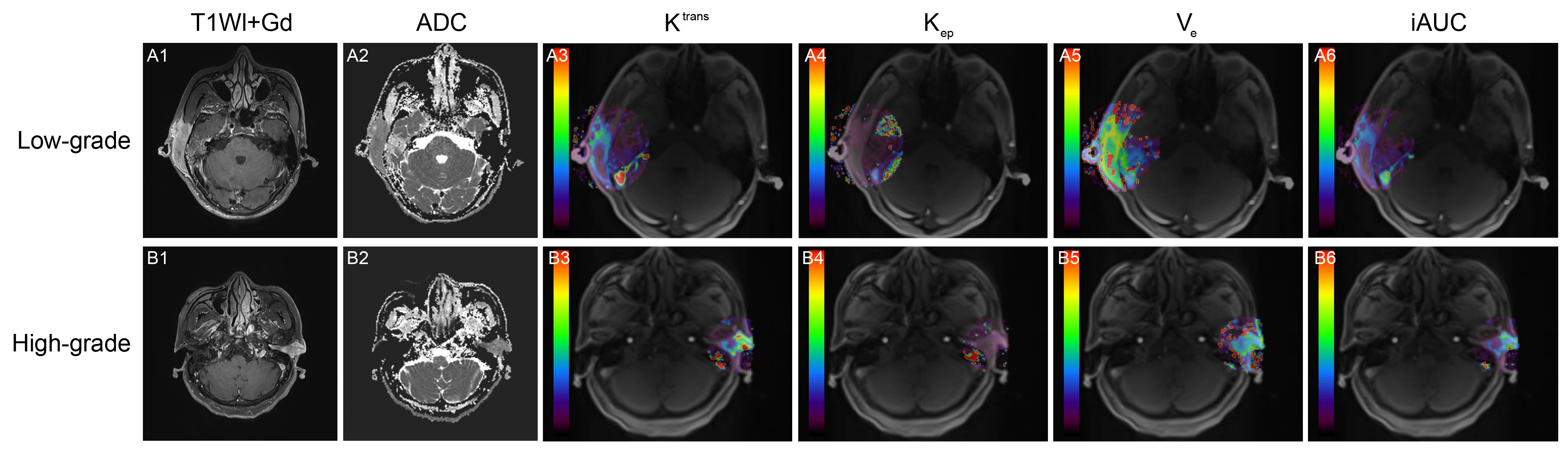
Methods: Retrospective diagnosis of 23 ear and temporal ACC patients was confirmed based on surgical pathology from March 2020 to April 2024. All patients underwent routine MRI, DWI, and DCE-MRI scans within one week before surgery. The lesion ADC values and DCE-MRI perfusion parameters, including Ve, Kep, Ktrans, and iAUC, were measured. Consistency tests were conducted on the measurements done by two physicians. The ADC values and DCE-MRI perfusion parameters between different pathological grades were compared. The correlation among all parameters and ACC pathological grading were analyzed, and the diagnostic accuracy of each parameter was assessed using receiver-operating-characteristic (ROC) curves.
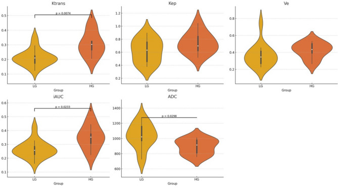
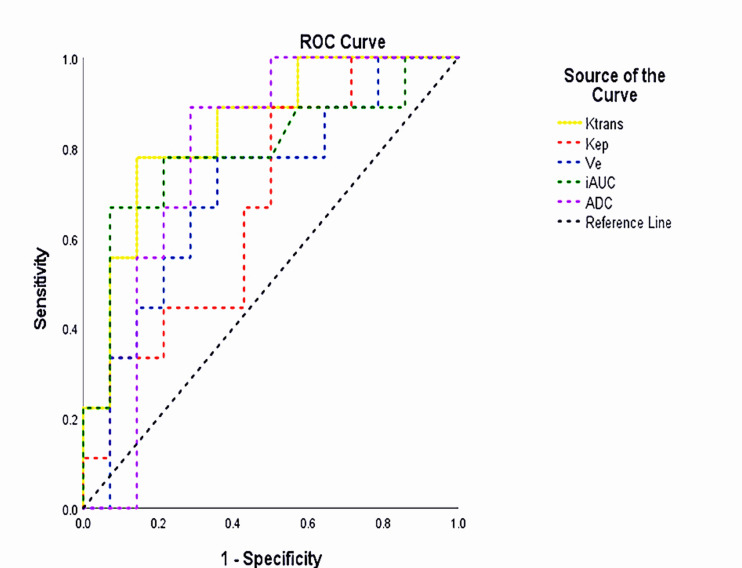
Results: The measurements from the two observers showed high consistency (ICC > 0.9). Ktrans, iAUC, and ADC values demonstrated significant differences between different pathological grades (P < .01, P < .05, P < .05). Correlation analysis indicated that Ktrans, Kep, Ve, and iAUC were positively correlated with ACC pathological grading, and Ktrans demonstrated the most robust correlation (correlation coefficient r = .578, P < .01). In contrast, ADC values were markedly and negatively correlated with pathological grading (r=-.470, P < .05). In ROC analysis, the area-under-the-curve (AUC) for Ktrans, iAUC, and ADC were 0.841, 0.790, and 0.778, respectively, all higher than those for Kep and Ve. Ktrans showed the best diagnostic performance.
Conclusion: Combining DCE-MRI perfusion parameters with ADC values provides a non-invasive and effective method for preoperative grading of ACC, with Ktrans, iAUC, and ADC showing strong diagnostic potential. These findings support more accurate tumor characterization and personalized treatment planning, warranting further validation in larger prospective studies.
期刊介绍:
BMC Medical Imaging is an open access journal publishing original peer-reviewed research articles in the development, evaluation, and use of imaging techniques and image processing tools to diagnose and manage disease.

 求助内容:
求助内容: 应助结果提醒方式:


