Heelim Lee, Jae Suk Jung, Jiwoo Lee, Seung Il Song, Jeong Keun Lee, Ji Min Kim, Bumhee Park, Inwhee Park, Suk Ji
{"title":"锥形束计算机断层扫描对慢性肾病患者下颌骨异常的评估:一项回顾性研究。","authors":"Heelim Lee, Jae Suk Jung, Jiwoo Lee, Seung Il Song, Jeong Keun Lee, Ji Min Kim, Bumhee Park, Inwhee Park, Suk Ji","doi":"10.2340/aos.v84.44619","DOIUrl":null,"url":null,"abstract":"<p><strong>Objective: </strong>This study evaluated mandibular cortical thickness and morphological changes in chronic kidney disease (CKD) patients using cone beam computed tomography (CBCT) and their correlation with bone metabolism markers.</p><p><strong>Methods: </strong>CKD patients were divided into CKD-I (eGFR (estimated glomerular filtration rate) < 30) and CKD-II (eGFR ≥ 30) groups, with healthy controls for comparison. Mental index (MI), antegonial index (AI), and panoramic mandibular index (PMI) were compared using Kruskal-Wallis test. Mandibular cortical index (MCI) classifications, lamina dura loss, and soft tissue calcifications were assessed using Fisher's test. Relationships between serum bone metabolism markers and radiomorphometric indices were analyzed by linear regression.</p><p><strong>Results: </strong>The study included 94 CKD patients (56 CKD-I, 38 CKD-II) and 88 controls. MI and AI were significantly lower in CKD-I versus controls (p < 0.05). MCI class I was less prevalent in CKD-I (5.4%) than controls (35.2%) (p < 0.001). Lamina dura loss (p = 0.006) and soft tissue calcifications (p = 0.009) occurred more frequently in CKD groups. Elevated alkaline phosphatase was associated with reduced cortical thickness (p < 0.01).</p><p><strong>Conclusions: </strong>Although findings should be interpreted considering the retrospective design's limitations, CBCT revealed significant bone abnormalities in CKD patients, with compromised bone quality and reduced mandibular cortical thickness, especially in advanced renal impairment, suggesting its value for pre-implant bone quality assessment in CKD patients.</p>","PeriodicalId":7313,"journal":{"name":"Acta Odontologica Scandinavica","volume":"84 ","pages":"517-526"},"PeriodicalIF":1.9000,"publicationDate":"2025-09-03","publicationTypes":"Journal Article","fieldsOfStudy":null,"isOpenAccess":false,"openAccessPdf":"https://www.ncbi.nlm.nih.gov/pmc/articles/PMC12416334/pdf/","citationCount":"0","resultStr":"{\"title\":\"Evaluation of mandibular bone abnormalities in patients with chronic kidney disease using cone beam computed tomography: A retrospective study.\",\"authors\":\"Heelim Lee, Jae Suk Jung, Jiwoo Lee, Seung Il Song, Jeong Keun Lee, Ji Min Kim, Bumhee Park, Inwhee Park, Suk Ji\",\"doi\":\"10.2340/aos.v84.44619\",\"DOIUrl\":null,\"url\":null,\"abstract\":\"<p><strong>Objective: </strong>This study evaluated mandibular cortical thickness and morphological changes in chronic kidney disease (CKD) patients using cone beam computed tomography (CBCT) and their correlation with bone metabolism markers.</p><p><strong>Methods: </strong>CKD patients were divided into CKD-I (eGFR (estimated glomerular filtration rate) < 30) and CKD-II (eGFR ≥ 30) groups, with healthy controls for comparison. Mental index (MI), antegonial index (AI), and panoramic mandibular index (PMI) were compared using Kruskal-Wallis test. Mandibular cortical index (MCI) classifications, lamina dura loss, and soft tissue calcifications were assessed using Fisher's test. Relationships between serum bone metabolism markers and radiomorphometric indices were analyzed by linear regression.</p><p><strong>Results: </strong>The study included 94 CKD patients (56 CKD-I, 38 CKD-II) and 88 controls. MI and AI were significantly lower in CKD-I versus controls (p < 0.05). MCI class I was less prevalent in CKD-I (5.4%) than controls (35.2%) (p < 0.001). Lamina dura loss (p = 0.006) and soft tissue calcifications (p = 0.009) occurred more frequently in CKD groups. Elevated alkaline phosphatase was associated with reduced cortical thickness (p < 0.01).</p><p><strong>Conclusions: </strong>Although findings should be interpreted considering the retrospective design's limitations, CBCT revealed significant bone abnormalities in CKD patients, with compromised bone quality and reduced mandibular cortical thickness, especially in advanced renal impairment, suggesting its value for pre-implant bone quality assessment in CKD patients.</p>\",\"PeriodicalId\":7313,\"journal\":{\"name\":\"Acta Odontologica Scandinavica\",\"volume\":\"84 \",\"pages\":\"517-526\"},\"PeriodicalIF\":1.9000,\"publicationDate\":\"2025-09-03\",\"publicationTypes\":\"Journal Article\",\"fieldsOfStudy\":null,\"isOpenAccess\":false,\"openAccessPdf\":\"https://www.ncbi.nlm.nih.gov/pmc/articles/PMC12416334/pdf/\",\"citationCount\":\"0\",\"resultStr\":null,\"platform\":\"Semanticscholar\",\"paperid\":null,\"PeriodicalName\":\"Acta Odontologica Scandinavica\",\"FirstCategoryId\":\"3\",\"ListUrlMain\":\"https://doi.org/10.2340/aos.v84.44619\",\"RegionNum\":4,\"RegionCategory\":\"医学\",\"ArticlePicture\":[],\"TitleCN\":null,\"AbstractTextCN\":null,\"PMCID\":null,\"EPubDate\":\"\",\"PubModel\":\"\",\"JCR\":\"Q3\",\"JCRName\":\"DENTISTRY, ORAL SURGERY & MEDICINE\",\"Score\":null,\"Total\":0}","platform":"Semanticscholar","paperid":null,"PeriodicalName":"Acta Odontologica Scandinavica","FirstCategoryId":"3","ListUrlMain":"https://doi.org/10.2340/aos.v84.44619","RegionNum":4,"RegionCategory":"医学","ArticlePicture":[],"TitleCN":null,"AbstractTextCN":null,"PMCID":null,"EPubDate":"","PubModel":"","JCR":"Q3","JCRName":"DENTISTRY, ORAL SURGERY & MEDICINE","Score":null,"Total":0}
Evaluation of mandibular bone abnormalities in patients with chronic kidney disease using cone beam computed tomography: A retrospective study.
Objective: This study evaluated mandibular cortical thickness and morphological changes in chronic kidney disease (CKD) patients using cone beam computed tomography (CBCT) and their correlation with bone metabolism markers.
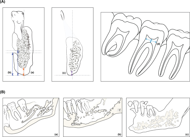
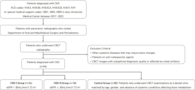
Methods: CKD patients were divided into CKD-I (eGFR (estimated glomerular filtration rate) < 30) and CKD-II (eGFR ≥ 30) groups, with healthy controls for comparison. Mental index (MI), antegonial index (AI), and panoramic mandibular index (PMI) were compared using Kruskal-Wallis test. Mandibular cortical index (MCI) classifications, lamina dura loss, and soft tissue calcifications were assessed using Fisher's test. Relationships between serum bone metabolism markers and radiomorphometric indices were analyzed by linear regression.
Results: The study included 94 CKD patients (56 CKD-I, 38 CKD-II) and 88 controls. MI and AI were significantly lower in CKD-I versus controls (p < 0.05). MCI class I was less prevalent in CKD-I (5.4%) than controls (35.2%) (p < 0.001). Lamina dura loss (p = 0.006) and soft tissue calcifications (p = 0.009) occurred more frequently in CKD groups. Elevated alkaline phosphatase was associated with reduced cortical thickness (p < 0.01).
Conclusions: Although findings should be interpreted considering the retrospective design's limitations, CBCT revealed significant bone abnormalities in CKD patients, with compromised bone quality and reduced mandibular cortical thickness, especially in advanced renal impairment, suggesting its value for pre-implant bone quality assessment in CKD patients.
 求助内容:
求助内容: 应助结果提醒方式:


