报告腹部扫描的ADPK疾病表型
IF 5.7
2区 医学
Q1 UROLOGY & NEPHROLOGY
引用次数: 0
摘要
腹部磁共振成像(MRI)和计算机断层扫描(CT)扫描的肾脏和肝脏体积是肾脏疾病:改善总体结局(KDIGO)推荐的常染色体显性多囊肾病(ADPKD)进展和治疗反应的关键生物标志物。本研究的目的是确定这些生物标志物在放射学报告中出现的频率及其可重复性。方法由2名观察员对腹部外MRI (n = 102)和CT (n = 43)研究进行回顾性和独立的回顾,以了解报告adpkd相关发现的患病率,并由2名放射科医生解决差异。这两名放射科医生独立地重新评估所有检查,以评估观察者之间的可重复性。结果来自88家机构的122名放射科医生的外部报告(n = 145,男性:46%,中位年龄:47[四分位数范围:35-61]岁)中只有30份(21%)报告包含肾脏体积。在140个包括整个肝脏的影像学检查中,1个(1%)外部报告提供了肝脏体积。外部和研究放射科医生肾脏体积测量的比较显示,使用椭球法的绝对中位数差为15%,使用模型辅助轮廓法的绝对中位数差为8%。145份外部报告中未提及的其他阳性发现包括脐疝(n = 44)、肝脂肪变性(n = 3)、腹股沟疝(n = 4)、胰腺囊肿(n = 6)和严重下腔静脉(IVC)囊肿压迫(n = 2)。结论放射科医师对ADPKD的复杂囊肿、钙化、腹水和腹主动脉瘤的报告可靠。然而,通过纳入肾脏体积和临床指南推荐的ADPKD的其他重要影像学特征,这些报告的临床效用可以得到更可靠的改善。本文章由计算机程序翻译,如有差异,请以英文原文为准。
Reporting ADPK Disease Phenotypes on Abdominal Scans
Introduction
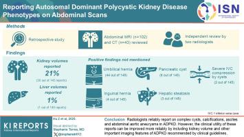
Kidney and liver volumes from abdominal magnetic resonance imaging (MRI) and computed tomography (CT) scans are critical biomarkers recommended by Kidney Disease: Improving Global Outcomes (KDIGO) for autosomal dominant polycystic kidney disease (ADPKD) progression and response to therapy. The purpose of this study was to determine how often these biomarkers are included in radiology reports as well as their reproducibility.
Methods
Outside abdominal MRI (n = 102) and CT (n = 43) studies were reviewed retrospectively and independently by 2 observers for prevalence of reporting ADPKD-relevant findings with discrepancies resolved by 2 radiologists. These 2 radiologists independently reevaluated all examinations to assess interobserver reproducibility.
Results
Outside reports (n = 145; males: 46%; median age: 47 [interquartile range: 35–61] years) by 122 radiologists from 88 institutions included kidney volumes in only 30 (21%) reports. Out of 140 imaging examinations that included the entire liver, 1 (1%) outside report provided liver volume. Comparison of outside and study radiologists’ kidney volume measurements showed a median absolute difference of 15%, using the ellipsoidal method and 8% using model-assisted contouring. Additional positive findings not mentioned in 145 outside reports included umbilical hernia (n = 44), hepatic steatosis (n = 3), inguinal hernia (n = 4), pancreatic cyst (n = 6) and severe inferior vena cava (IVC) compression by cysts (n = 2).
Conclusion
Radiologists report reliably on complex cysts, calcifications, ascites, and abdominal aortic aneurysms in ADPKD. However, the clinical utility of these reports can be improved more reliably by including kidney volume and other important imaging features of ADPKD recommended by clinical guidelines.
求助全文
通过发布文献求助,成功后即可免费获取论文全文。
去求助
来源期刊
Kidney International Reports
Medicine-Nephrology
CiteScore
7.70
自引率
3.30%
发文量
1578
审稿时长
8 weeks
期刊介绍:
Kidney International Reports, an official journal of the International Society of Nephrology, is a peer-reviewed, open access journal devoted to the publication of leading research and developments related to kidney disease. With the primary aim of contributing to improved care of patients with kidney disease, the journal will publish original clinical and select translational articles and educational content related to the pathogenesis, evaluation and management of acute and chronic kidney disease, end stage renal disease (including transplantation), acid-base, fluid and electrolyte disturbances and hypertension. Of particular interest are submissions related to clinical trials, epidemiology, systematic reviews (including meta-analyses) and outcomes research. The journal will also provide a platform for wider dissemination of national and regional guidelines as well as consensus meeting reports.
 求助内容:
求助内容: 应助结果提醒方式:


