Zohar Eyal, Rachael Deis, Anna Gorelick-Ashkenazi, Yuval Barzilay, Yonatan Broder, Asher Perry Kellum, Neta Varsano, Michal Hartstein, Andrea Sorrentino, Ron Rotkopf, Ifat Kaplan-Ashiri, Katya Rechav, Rebecca Metzler, Lothar Houben, Leeor Kronik, Peter Rez, Dvir Gur
{"title":"pH值的变化使鸟嘌呤晶体在虹膜小体内形成","authors":"Zohar Eyal, Rachael Deis, Anna Gorelick-Ashkenazi, Yuval Barzilay, Yonatan Broder, Asher Perry Kellum, Neta Varsano, Michal Hartstein, Andrea Sorrentino, Ron Rotkopf, Ifat Kaplan-Ashiri, Katya Rechav, Rebecca Metzler, Lothar Houben, Leeor Kronik, Peter Rez, Dvir Gur","doi":"10.1038/s41589-025-02020-0","DOIUrl":null,"url":null,"abstract":"<p>Many animals produce vivid colors by reflecting and amplifying light with stacked guanine crystals within membrane-bound organelles called iridosomes. While the presence of guanine crystals in iridosomes is well documented, the mechanisms facilitating the accumulation of water-insoluble guanine and driving its crystallization remain unclear. Here we used cryo-electron microscopy, live-cell pH imaging, pharmacological perturbations and spectroscopy to study iridosome maturation in zebrafish. Cryo-electron and synchrotron-based soft X-ray microscopies revealed that amorphous guanine initially accumulates in early-stage iridosomes in its protonated state. Live-cell imaging with a pH sensor demonstrated that early iridosomes are acidic, with pH gradually neutralizing during development. Inhibiting V-ATPase disrupted this acidification and significantly reduced crystal formation, indicating its role in pH regulation. Our findings reveal insights into the molecular mechanisms facilitating guanine formation within iridosomes, emphasizing the pivotal role of pH alternations in the precise formation of biogenic crystals.</p><figure></figure>","PeriodicalId":18832,"journal":{"name":"Nature chemical biology","volume":"58 1","pages":""},"PeriodicalIF":13.7000,"publicationDate":"2025-09-02","publicationTypes":"Journal Article","fieldsOfStudy":null,"isOpenAccess":false,"openAccessPdf":"","citationCount":"0","resultStr":"{\"title\":\"pH variations enable guanine crystal formation within iridosomes\",\"authors\":\"Zohar Eyal, Rachael Deis, Anna Gorelick-Ashkenazi, Yuval Barzilay, Yonatan Broder, Asher Perry Kellum, Neta Varsano, Michal Hartstein, Andrea Sorrentino, Ron Rotkopf, Ifat Kaplan-Ashiri, Katya Rechav, Rebecca Metzler, Lothar Houben, Leeor Kronik, Peter Rez, Dvir Gur\",\"doi\":\"10.1038/s41589-025-02020-0\",\"DOIUrl\":null,\"url\":null,\"abstract\":\"<p>Many animals produce vivid colors by reflecting and amplifying light with stacked guanine crystals within membrane-bound organelles called iridosomes. While the presence of guanine crystals in iridosomes is well documented, the mechanisms facilitating the accumulation of water-insoluble guanine and driving its crystallization remain unclear. Here we used cryo-electron microscopy, live-cell pH imaging, pharmacological perturbations and spectroscopy to study iridosome maturation in zebrafish. Cryo-electron and synchrotron-based soft X-ray microscopies revealed that amorphous guanine initially accumulates in early-stage iridosomes in its protonated state. Live-cell imaging with a pH sensor demonstrated that early iridosomes are acidic, with pH gradually neutralizing during development. Inhibiting V-ATPase disrupted this acidification and significantly reduced crystal formation, indicating its role in pH regulation. Our findings reveal insights into the molecular mechanisms facilitating guanine formation within iridosomes, emphasizing the pivotal role of pH alternations in the precise formation of biogenic crystals.</p><figure></figure>\",\"PeriodicalId\":18832,\"journal\":{\"name\":\"Nature chemical biology\",\"volume\":\"58 1\",\"pages\":\"\"},\"PeriodicalIF\":13.7000,\"publicationDate\":\"2025-09-02\",\"publicationTypes\":\"Journal Article\",\"fieldsOfStudy\":null,\"isOpenAccess\":false,\"openAccessPdf\":\"\",\"citationCount\":\"0\",\"resultStr\":null,\"platform\":\"Semanticscholar\",\"paperid\":null,\"PeriodicalName\":\"Nature chemical biology\",\"FirstCategoryId\":\"99\",\"ListUrlMain\":\"https://doi.org/10.1038/s41589-025-02020-0\",\"RegionNum\":1,\"RegionCategory\":\"生物学\",\"ArticlePicture\":[],\"TitleCN\":null,\"AbstractTextCN\":null,\"PMCID\":null,\"EPubDate\":\"\",\"PubModel\":\"\",\"JCR\":\"Q1\",\"JCRName\":\"BIOCHEMISTRY & MOLECULAR BIOLOGY\",\"Score\":null,\"Total\":0}","platform":"Semanticscholar","paperid":null,"PeriodicalName":"Nature chemical biology","FirstCategoryId":"99","ListUrlMain":"https://doi.org/10.1038/s41589-025-02020-0","RegionNum":1,"RegionCategory":"生物学","ArticlePicture":[],"TitleCN":null,"AbstractTextCN":null,"PMCID":null,"EPubDate":"","PubModel":"","JCR":"Q1","JCRName":"BIOCHEMISTRY & MOLECULAR BIOLOGY","Score":null,"Total":0}
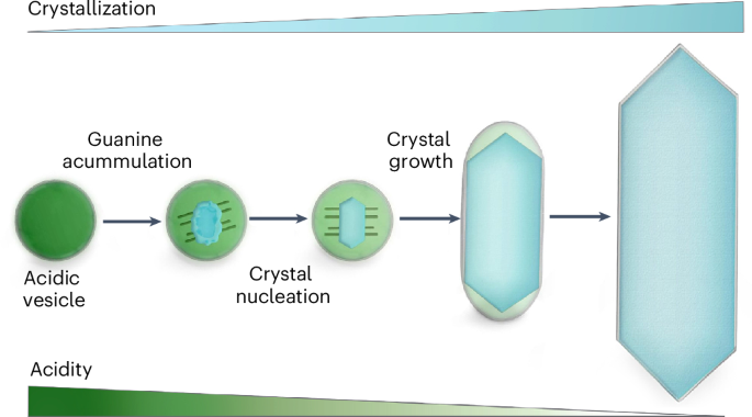
pH variations enable guanine crystal formation within iridosomes
Many animals produce vivid colors by reflecting and amplifying light with stacked guanine crystals within membrane-bound organelles called iridosomes. While the presence of guanine crystals in iridosomes is well documented, the mechanisms facilitating the accumulation of water-insoluble guanine and driving its crystallization remain unclear. Here we used cryo-electron microscopy, live-cell pH imaging, pharmacological perturbations and spectroscopy to study iridosome maturation in zebrafish. Cryo-electron and synchrotron-based soft X-ray microscopies revealed that amorphous guanine initially accumulates in early-stage iridosomes in its protonated state. Live-cell imaging with a pH sensor demonstrated that early iridosomes are acidic, with pH gradually neutralizing during development. Inhibiting V-ATPase disrupted this acidification and significantly reduced crystal formation, indicating its role in pH regulation. Our findings reveal insights into the molecular mechanisms facilitating guanine formation within iridosomes, emphasizing the pivotal role of pH alternations in the precise formation of biogenic crystals.
期刊介绍:
Nature Chemical Biology stands as an esteemed international monthly journal, offering a prominent platform for the chemical biology community to showcase top-tier original research and commentary. Operating at the crossroads of chemistry, biology, and related disciplines, chemical biology utilizes scientific ideas and approaches to comprehend and manipulate biological systems with molecular precision.
The journal embraces contributions from the growing community of chemical biologists, encompassing insights from chemists applying principles and tools to biological inquiries and biologists striving to comprehend and control molecular-level biological processes. We prioritize studies unveiling significant conceptual or practical advancements in areas where chemistry and biology intersect, emphasizing basic research, especially those reporting novel chemical or biological tools and offering profound molecular-level insights into underlying biological mechanisms.
Nature Chemical Biology also welcomes manuscripts describing applied molecular studies at the chemistry-biology interface due to the broad utility of chemical biology approaches in manipulating or engineering biological systems. Irrespective of scientific focus, we actively seek submissions that creatively blend chemistry and biology, particularly those providing substantial conceptual or methodological breakthroughs with the potential to open innovative research avenues. The journal maintains a robust and impartial review process, emphasizing thorough chemical and biological characterization.
 求助内容:
求助内容: 应助结果提醒方式:


