氮芥会导致进行性组织损伤,严重的DNA损伤和角膜坏死
IF 2.7
2区 医学
Q1 OPHTHALMOLOGY
引用次数: 0
摘要
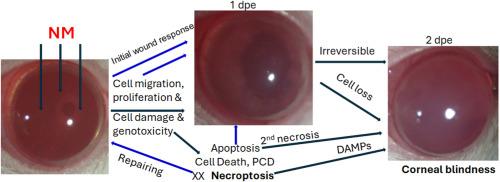
眼睛很敏感,容易受到硫和氮芥(NM)等发泡剂的伤害。为了评估纳米暴露对眼表的不良影响,我们使用猪和大鼠的角膜和纳米湿润滤液纸盘来评估纳米对角膜健康的离体和体内影响。在培养的猪角膜(N = 5)中,5mg /ml NM引起暴露时间依赖性损伤,其进展时间为NM暴露后1至2天(dpe),程序性细胞死亡增加,特别是在2 dpe时坏死。在大鼠角膜(N = 4)中,5mg /mL NM引起角膜混浊、点状角膜炎、瞳孔异角、角膜敏感性降低。在1-dpe的组织水平上,上皮细胞表现出前缘,前排上皮细胞主动迁移覆盖创面,顶端层c-Casp3阳性,基底和/或翼细胞p-RIPK3阳性。大多数角膜细胞γ - h2ax阳性,基质细胞tunel阳性,但c- casp3或p- ripk3阴性。在2-dpe时,只有少数细胞c-Casp3阳性,而大多数角膜顶侧细胞p-RIPK3阳性。这些变化与纳米暴露大鼠角膜基底膜破裂和紧密连接丧失有关。最后,在培养的猪角膜中,抑制坏死下垂可防止NM引起的角膜组织破坏。我们的数据表明纳米诱导的角膜损伤的严重程度可能与坏死下垂的增加有关,靶向坏死下垂可以减少或预防纳米暴露引起的角膜恶化。本文章由计算机程序翻译,如有差异,请以英文原文为准。
Nitrogen mustard causes progressive tissue injury, severe DNA damage, and necroptosis in the cornea
The eye is sensitive and vulnerable to vesicants such as sulfur and nitrogen mustard (NM). To assess the adverse effects of NM exposure on the ocular surface, we used porcine and rat corneas and NM-wetted filtrate paper disks to assess NM's ex vivo and in vivo effects on corneal health. In cultured porcine corneas (N = 5), 5 mg/ml NM caused exposure time-dependent damage, which progressed from 1 to 2 days post-NM exposure (dpe), with increased programmed cell death, particularly necroptosis at 2 dpe. In rat corneas (N = 4), 5 mg/mL NM caused corneal opacification, punctate keratitis, pupil anisocoria, and decreased corneal sensitivity. At the tissue level at 1-dpe, epithelial cells exhibited a leading edge, the front row of epithelial cells that actively migrate to cover the wound area, with apical layers being c-Casp3 positive and basal and/or wing cells being p-RIPK3 positive. Most corneal cells were γH2AX-positive, and stromal cells were TUNEL-positive but c-Casp3-or p-RIPK3-negative. At 2-dpe, only a few cells were c-Casp3 positive, while most cells on the apical side of the cornea were p-RIPK3 positive. These changes were associated with basement membrane breakdown and tight junction loss in NM-exposed rat corneas. Finally, in cultured porcine corneas, the inhibition of necroptosis resulted in the prevention of corneal tissue destruction caused by NM. Our data suggest that the severity of NM-induced corneal injuries may be linked to increased necroptosis, and targeting necroptosis may reduce or prevent corneal deterioration caused by NM exposure.
求助全文
通过发布文献求助,成功后即可免费获取论文全文。
去求助
来源期刊

Experimental eye research
医学-眼科学
CiteScore
6.80
自引率
5.90%
发文量
323
审稿时长
66 days
期刊介绍:
The primary goal of Experimental Eye Research is to publish original research papers on all aspects of experimental biology of the eye and ocular tissues that seek to define the mechanisms of normal function and/or disease. Studies of ocular tissues that encompass the disciplines of cell biology, developmental biology, genetics, molecular biology, physiology, biochemistry, biophysics, immunology or microbiology are most welcomed. Manuscripts that are purely clinical or in a surgical area of ophthalmology are not appropriate for submission to Experimental Eye Research and if received will be returned without review.
 求助内容:
求助内容: 应助结果提醒方式:


