{"title":"应用MpMRI和18f - psma - pet /CT预测前列腺癌前列腺外展的多模态成像深度学习模型","authors":"Fei Yao, Heng Lin, Ying-Nan Xue, Yuan-Di Zhuang, Shu-Ying Bian, Ya-Yun Zhang, Yun-Jun Yang, Ke-Hua Pan","doi":"10.1186/s40644-025-00927-4","DOIUrl":null,"url":null,"abstract":"<p><strong>Objective: </strong>This study aimed to construct a multimodal imaging deep learning (DL) model integrating mpMRI and <sup>18</sup>F-PSMA-PET/CT for the prediction of extraprostatic extension (EPE) in prostate cancer, and to assess its effectiveness in enhancing the diagnostic accuracy of radiologists.</p><p><strong>Methods: </strong>Clinical and imaging data were retrospectively collected from patients with pathologically confirmed prostate cancer (PCa) who underwent radical prostatectomy (RP). Data were collected from a primary institution (Center 1, n = 197) between January 2019 and June 2022 and an external institution (Center 2, n = 36) between July 2021 and November 2022. A multimodal DL model incorporating mpMRI and <sup>18</sup>F-PSMA-PET/CT was developed to support radiologists in assessing EPE using the EPE-grade scoring system. The predictive performance of the DL model was compared with that of single-modality models, as well as with radiologist assessments with and without model assistance. Clinical net benefit of the model was also assessed.</p><p><strong>Results: </strong>For patients in Center 1, the area under the curve (AUC) for predicting EPE was 0.76 (0.72-0.80), 0.77 (0.70-0.82), and 0.82 (0.78-0.87) for the mpMRI-based DL model, PET/CT-based DL model, and the combined mpMRI + PET/CT multimodal DL model, respectively. In the external test set (Center 2), the AUCs for these models were 0.75 (0.60-0.88), 0.77 (0.72-0.88), and 0.81 (0.63-0.97), respectively. The multimodal DL model demonstrated superior predictive accuracy compared to single-modality models in both internal and external validations. The deep learning-assisted EPE-grade scoring model significantly improved AUC and sensitivity compared to radiologist EPE-grade scoring alone (P < 0.05), with a modest reduction in specificity. Additionally, the deep learning-assisted scoring model provided greater clinical net benefit than the radiologist EPE-grade score used by radiologists alone.</p><p><strong>Conclusion: </strong>The multimodal imaging deep learning model, integrating mpMRI and 18 F-PSMA PET/CT, demonstrates promising predictive performance for EPE in prostate cancer and enhances the accuracy of radiologists in EPE assessment. The model holds potential as a supportive tool for more individualized and precise therapeutic decision-making.</p>","PeriodicalId":9548,"journal":{"name":"Cancer Imaging","volume":"25 1","pages":"103"},"PeriodicalIF":3.5000,"publicationDate":"2025-08-19","publicationTypes":"Journal Article","fieldsOfStudy":null,"isOpenAccess":false,"openAccessPdf":"https://www.ncbi.nlm.nih.gov/pmc/articles/PMC12366157/pdf/","citationCount":"0","resultStr":"{\"title\":\"Multimodal imaging deep learning model for predicting extraprostatic extension in prostate cancer using MpMRI and 18 F-PSMA-PET/CT.\",\"authors\":\"Fei Yao, Heng Lin, Ying-Nan Xue, Yuan-Di Zhuang, Shu-Ying Bian, Ya-Yun Zhang, Yun-Jun Yang, Ke-Hua Pan\",\"doi\":\"10.1186/s40644-025-00927-4\",\"DOIUrl\":null,\"url\":null,\"abstract\":\"<p><strong>Objective: </strong>This study aimed to construct a multimodal imaging deep learning (DL) model integrating mpMRI and <sup>18</sup>F-PSMA-PET/CT for the prediction of extraprostatic extension (EPE) in prostate cancer, and to assess its effectiveness in enhancing the diagnostic accuracy of radiologists.</p><p><strong>Methods: </strong>Clinical and imaging data were retrospectively collected from patients with pathologically confirmed prostate cancer (PCa) who underwent radical prostatectomy (RP). Data were collected from a primary institution (Center 1, n = 197) between January 2019 and June 2022 and an external institution (Center 2, n = 36) between July 2021 and November 2022. A multimodal DL model incorporating mpMRI and <sup>18</sup>F-PSMA-PET/CT was developed to support radiologists in assessing EPE using the EPE-grade scoring system. The predictive performance of the DL model was compared with that of single-modality models, as well as with radiologist assessments with and without model assistance. Clinical net benefit of the model was also assessed.</p><p><strong>Results: </strong>For patients in Center 1, the area under the curve (AUC) for predicting EPE was 0.76 (0.72-0.80), 0.77 (0.70-0.82), and 0.82 (0.78-0.87) for the mpMRI-based DL model, PET/CT-based DL model, and the combined mpMRI + PET/CT multimodal DL model, respectively. In the external test set (Center 2), the AUCs for these models were 0.75 (0.60-0.88), 0.77 (0.72-0.88), and 0.81 (0.63-0.97), respectively. The multimodal DL model demonstrated superior predictive accuracy compared to single-modality models in both internal and external validations. The deep learning-assisted EPE-grade scoring model significantly improved AUC and sensitivity compared to radiologist EPE-grade scoring alone (P < 0.05), with a modest reduction in specificity. Additionally, the deep learning-assisted scoring model provided greater clinical net benefit than the radiologist EPE-grade score used by radiologists alone.</p><p><strong>Conclusion: </strong>The multimodal imaging deep learning model, integrating mpMRI and 18 F-PSMA PET/CT, demonstrates promising predictive performance for EPE in prostate cancer and enhances the accuracy of radiologists in EPE assessment. The model holds potential as a supportive tool for more individualized and precise therapeutic decision-making.</p>\",\"PeriodicalId\":9548,\"journal\":{\"name\":\"Cancer Imaging\",\"volume\":\"25 1\",\"pages\":\"103\"},\"PeriodicalIF\":3.5000,\"publicationDate\":\"2025-08-19\",\"publicationTypes\":\"Journal Article\",\"fieldsOfStudy\":null,\"isOpenAccess\":false,\"openAccessPdf\":\"https://www.ncbi.nlm.nih.gov/pmc/articles/PMC12366157/pdf/\",\"citationCount\":\"0\",\"resultStr\":null,\"platform\":\"Semanticscholar\",\"paperid\":null,\"PeriodicalName\":\"Cancer Imaging\",\"FirstCategoryId\":\"3\",\"ListUrlMain\":\"https://doi.org/10.1186/s40644-025-00927-4\",\"RegionNum\":2,\"RegionCategory\":\"医学\",\"ArticlePicture\":[],\"TitleCN\":null,\"AbstractTextCN\":null,\"PMCID\":null,\"EPubDate\":\"\",\"PubModel\":\"\",\"JCR\":\"Q2\",\"JCRName\":\"ONCOLOGY\",\"Score\":null,\"Total\":0}","platform":"Semanticscholar","paperid":null,"PeriodicalName":"Cancer Imaging","FirstCategoryId":"3","ListUrlMain":"https://doi.org/10.1186/s40644-025-00927-4","RegionNum":2,"RegionCategory":"医学","ArticlePicture":[],"TitleCN":null,"AbstractTextCN":null,"PMCID":null,"EPubDate":"","PubModel":"","JCR":"Q2","JCRName":"ONCOLOGY","Score":null,"Total":0}
Multimodal imaging deep learning model for predicting extraprostatic extension in prostate cancer using MpMRI and 18 F-PSMA-PET/CT.
Objective: This study aimed to construct a multimodal imaging deep learning (DL) model integrating mpMRI and 18F-PSMA-PET/CT for the prediction of extraprostatic extension (EPE) in prostate cancer, and to assess its effectiveness in enhancing the diagnostic accuracy of radiologists.
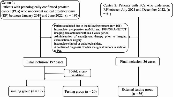
Methods: Clinical and imaging data were retrospectively collected from patients with pathologically confirmed prostate cancer (PCa) who underwent radical prostatectomy (RP). Data were collected from a primary institution (Center 1, n = 197) between January 2019 and June 2022 and an external institution (Center 2, n = 36) between July 2021 and November 2022. A multimodal DL model incorporating mpMRI and 18F-PSMA-PET/CT was developed to support radiologists in assessing EPE using the EPE-grade scoring system. The predictive performance of the DL model was compared with that of single-modality models, as well as with radiologist assessments with and without model assistance. Clinical net benefit of the model was also assessed.
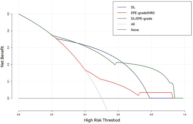
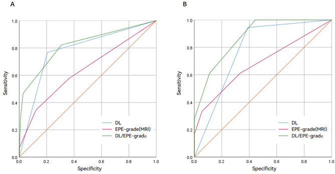
Results: For patients in Center 1, the area under the curve (AUC) for predicting EPE was 0.76 (0.72-0.80), 0.77 (0.70-0.82), and 0.82 (0.78-0.87) for the mpMRI-based DL model, PET/CT-based DL model, and the combined mpMRI + PET/CT multimodal DL model, respectively. In the external test set (Center 2), the AUCs for these models were 0.75 (0.60-0.88), 0.77 (0.72-0.88), and 0.81 (0.63-0.97), respectively. The multimodal DL model demonstrated superior predictive accuracy compared to single-modality models in both internal and external validations. The deep learning-assisted EPE-grade scoring model significantly improved AUC and sensitivity compared to radiologist EPE-grade scoring alone (P < 0.05), with a modest reduction in specificity. Additionally, the deep learning-assisted scoring model provided greater clinical net benefit than the radiologist EPE-grade score used by radiologists alone.
Conclusion: The multimodal imaging deep learning model, integrating mpMRI and 18 F-PSMA PET/CT, demonstrates promising predictive performance for EPE in prostate cancer and enhances the accuracy of radiologists in EPE assessment. The model holds potential as a supportive tool for more individualized and precise therapeutic decision-making.
Cancer ImagingONCOLOGY-RADIOLOGY, NUCLEAR MEDICINE & MEDICAL IMAGING
CiteScore
7.00
自引率
0.00%
发文量
66
审稿时长
>12 weeks
期刊介绍:
Cancer Imaging is an open access, peer-reviewed journal publishing original articles, reviews and editorials written by expert international radiologists working in oncology.
The journal encompasses CT, MR, PET, ultrasound, radionuclide and multimodal imaging in all kinds of malignant tumours, plus new developments, techniques and innovations. Topics of interest include:
Breast Imaging
Chest
Complications of treatment
Ear, Nose & Throat
Gastrointestinal
Hepatobiliary & Pancreatic
Imaging biomarkers
Interventional
Lymphoma
Measurement of tumour response
Molecular functional imaging
Musculoskeletal
Neuro oncology
Nuclear Medicine
Paediatric.

 求助内容:
求助内容: 应助结果提醒方式:


