{"title":"术前胸肌指数预测非小细胞肺癌患者无远处转移生存:一项回顾性研究。","authors":"Zhihui Shi, Lin Wu, Dengke Jiang, Ruiling Yang, Rui Liao, Lizhu Liu, Ruimin You, Yanli Li, Xingxiang Dong, Dafu Zhang, Jing Wang, Xuewen Zhang, Xiaobo Chen, Zhenhui Li","doi":"10.1186/s12880-025-01873-0","DOIUrl":null,"url":null,"abstract":"<p><strong>Background: </strong>Thoracic muscles contribute to respiration, is a crucial indicator for assessing functional recovery following lung resection. However, there is a lack of research on the long-term prognostic value of pectoralis muscle.</p><p><strong>Methods: </strong>Consecutive patients who underwent curative-intent resection for stage I to IIIA NSCLC between 2013 and 2018 at a cancer center were retrospectively identified. The Cox proportional hazard model was employed to analyze the correlation between pectoralis muscle index (PMI) and survival, with subgroup analyses conducted to explore potential heterogeneity among different subgroups. Finally, the relative influence of each parameter was compared using a gradient boosting model (GBM).</p><p><strong>Results: </strong>A total of 2110 patients (median (IQR) age 59 (52, 66) years) were evaluated. Kaplan-Meier survival analysis showed that the recurrence-free survival (RFS) and distant metastasis-free survival (DMFS) rate of patients in the high PMI group were higher than those in the low PMI group, all with P < 0.001. In the multivariable analysis, low PMI is still associated with shorter RFS (HR = 1.34, 95% CI: (1.10, 1.62), P = 0.004), DMFS (HR = 1.35, 95% CI: (1.11, 1.65), P = 0.003), lung MFS (HR = 1.47, 95% CI: (1.19, 1.81), P < 0.001) and bone MFS (HR = 1.38, 95% CI: (1.11, 1.73), P = 0.004). These associations were consistent in subgroup analysis of different gender, age, tumor stage, histologic type, and surgical approach group.</p><p><strong>Conclusions: </strong>Low PMI is significantly associated with worse distant metastasis-free survival (DMFS) and recurrence-free survival (RFS) in non-small cell lung cancer (NSCLC) patients, supporting its utility in refining preoperative risk stratification. When CT imaging lacks L3-level coverage, PMI offers a viable alternative for assessing muscle quality.</p>","PeriodicalId":9020,"journal":{"name":"BMC Medical Imaging","volume":"25 1","pages":"335"},"PeriodicalIF":3.2000,"publicationDate":"2025-08-19","publicationTypes":"Journal Article","fieldsOfStudy":null,"isOpenAccess":false,"openAccessPdf":"https://www.ncbi.nlm.nih.gov/pmc/articles/PMC12362906/pdf/","citationCount":"0","resultStr":"{\"title\":\"Preoperative pectoralis muscle index predicts distant metastasis-free survival in non-small cell lung cancer patients: a retrospective study.\",\"authors\":\"Zhihui Shi, Lin Wu, Dengke Jiang, Ruiling Yang, Rui Liao, Lizhu Liu, Ruimin You, Yanli Li, Xingxiang Dong, Dafu Zhang, Jing Wang, Xuewen Zhang, Xiaobo Chen, Zhenhui Li\",\"doi\":\"10.1186/s12880-025-01873-0\",\"DOIUrl\":null,\"url\":null,\"abstract\":\"<p><strong>Background: </strong>Thoracic muscles contribute to respiration, is a crucial indicator for assessing functional recovery following lung resection. However, there is a lack of research on the long-term prognostic value of pectoralis muscle.</p><p><strong>Methods: </strong>Consecutive patients who underwent curative-intent resection for stage I to IIIA NSCLC between 2013 and 2018 at a cancer center were retrospectively identified. The Cox proportional hazard model was employed to analyze the correlation between pectoralis muscle index (PMI) and survival, with subgroup analyses conducted to explore potential heterogeneity among different subgroups. Finally, the relative influence of each parameter was compared using a gradient boosting model (GBM).</p><p><strong>Results: </strong>A total of 2110 patients (median (IQR) age 59 (52, 66) years) were evaluated. Kaplan-Meier survival analysis showed that the recurrence-free survival (RFS) and distant metastasis-free survival (DMFS) rate of patients in the high PMI group were higher than those in the low PMI group, all with P < 0.001. In the multivariable analysis, low PMI is still associated with shorter RFS (HR = 1.34, 95% CI: (1.10, 1.62), P = 0.004), DMFS (HR = 1.35, 95% CI: (1.11, 1.65), P = 0.003), lung MFS (HR = 1.47, 95% CI: (1.19, 1.81), P < 0.001) and bone MFS (HR = 1.38, 95% CI: (1.11, 1.73), P = 0.004). These associations were consistent in subgroup analysis of different gender, age, tumor stage, histologic type, and surgical approach group.</p><p><strong>Conclusions: </strong>Low PMI is significantly associated with worse distant metastasis-free survival (DMFS) and recurrence-free survival (RFS) in non-small cell lung cancer (NSCLC) patients, supporting its utility in refining preoperative risk stratification. When CT imaging lacks L3-level coverage, PMI offers a viable alternative for assessing muscle quality.</p>\",\"PeriodicalId\":9020,\"journal\":{\"name\":\"BMC Medical Imaging\",\"volume\":\"25 1\",\"pages\":\"335\"},\"PeriodicalIF\":3.2000,\"publicationDate\":\"2025-08-19\",\"publicationTypes\":\"Journal Article\",\"fieldsOfStudy\":null,\"isOpenAccess\":false,\"openAccessPdf\":\"https://www.ncbi.nlm.nih.gov/pmc/articles/PMC12362906/pdf/\",\"citationCount\":\"0\",\"resultStr\":null,\"platform\":\"Semanticscholar\",\"paperid\":null,\"PeriodicalName\":\"BMC Medical Imaging\",\"FirstCategoryId\":\"3\",\"ListUrlMain\":\"https://doi.org/10.1186/s12880-025-01873-0\",\"RegionNum\":3,\"RegionCategory\":\"医学\",\"ArticlePicture\":[],\"TitleCN\":null,\"AbstractTextCN\":null,\"PMCID\":null,\"EPubDate\":\"\",\"PubModel\":\"\",\"JCR\":\"Q2\",\"JCRName\":\"RADIOLOGY, NUCLEAR MEDICINE & MEDICAL IMAGING\",\"Score\":null,\"Total\":0}","platform":"Semanticscholar","paperid":null,"PeriodicalName":"BMC Medical Imaging","FirstCategoryId":"3","ListUrlMain":"https://doi.org/10.1186/s12880-025-01873-0","RegionNum":3,"RegionCategory":"医学","ArticlePicture":[],"TitleCN":null,"AbstractTextCN":null,"PMCID":null,"EPubDate":"","PubModel":"","JCR":"Q2","JCRName":"RADIOLOGY, NUCLEAR MEDICINE & MEDICAL IMAGING","Score":null,"Total":0}
Preoperative pectoralis muscle index predicts distant metastasis-free survival in non-small cell lung cancer patients: a retrospective study.
Background: Thoracic muscles contribute to respiration, is a crucial indicator for assessing functional recovery following lung resection. However, there is a lack of research on the long-term prognostic value of pectoralis muscle.
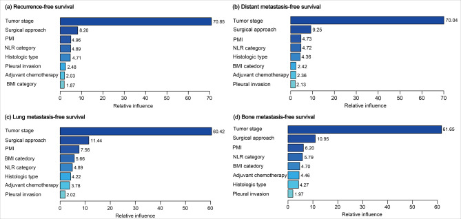
Methods: Consecutive patients who underwent curative-intent resection for stage I to IIIA NSCLC between 2013 and 2018 at a cancer center were retrospectively identified. The Cox proportional hazard model was employed to analyze the correlation between pectoralis muscle index (PMI) and survival, with subgroup analyses conducted to explore potential heterogeneity among different subgroups. Finally, the relative influence of each parameter was compared using a gradient boosting model (GBM).
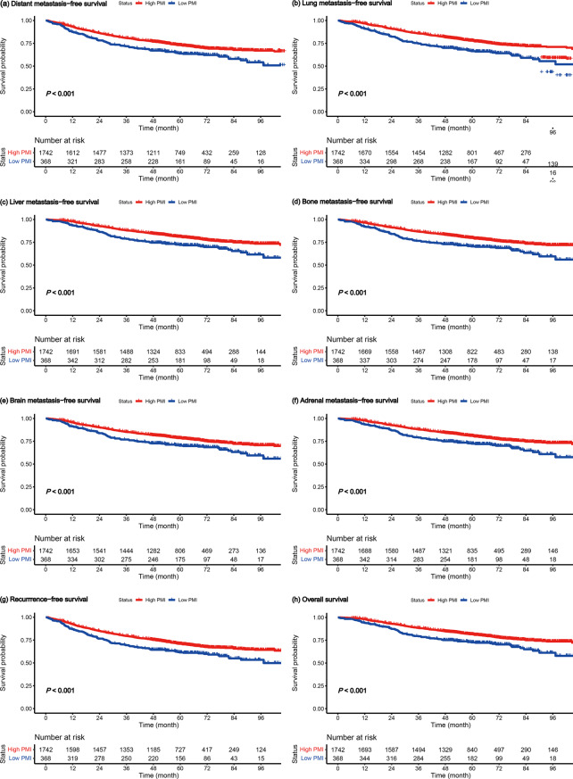
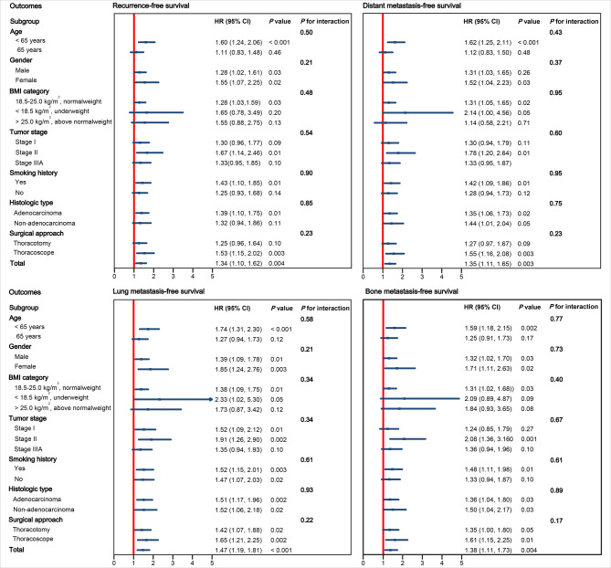
Results: A total of 2110 patients (median (IQR) age 59 (52, 66) years) were evaluated. Kaplan-Meier survival analysis showed that the recurrence-free survival (RFS) and distant metastasis-free survival (DMFS) rate of patients in the high PMI group were higher than those in the low PMI group, all with P < 0.001. In the multivariable analysis, low PMI is still associated with shorter RFS (HR = 1.34, 95% CI: (1.10, 1.62), P = 0.004), DMFS (HR = 1.35, 95% CI: (1.11, 1.65), P = 0.003), lung MFS (HR = 1.47, 95% CI: (1.19, 1.81), P < 0.001) and bone MFS (HR = 1.38, 95% CI: (1.11, 1.73), P = 0.004). These associations were consistent in subgroup analysis of different gender, age, tumor stage, histologic type, and surgical approach group.
Conclusions: Low PMI is significantly associated with worse distant metastasis-free survival (DMFS) and recurrence-free survival (RFS) in non-small cell lung cancer (NSCLC) patients, supporting its utility in refining preoperative risk stratification. When CT imaging lacks L3-level coverage, PMI offers a viable alternative for assessing muscle quality.
期刊介绍:
BMC Medical Imaging is an open access journal publishing original peer-reviewed research articles in the development, evaluation, and use of imaging techniques and image processing tools to diagnose and manage disease.

 求助内容:
求助内容: 应助结果提醒方式:


