{"title":"角膜缘放松切口联合超声乳化术与标准超声乳化术术后眼表组织损伤的比较。","authors":"Haiyan Wang, Xinlu Yu, Yanan Li, Lingjuan Sun, Mingqi Liu, Xiongwei Zhang, Ruibo Yang","doi":"10.2147/OPTH.S534006","DOIUrl":null,"url":null,"abstract":"<p><strong>Purpose: </strong>To investigate the effect of phacoemulsification combined with LRIs on the ocular surface at different postoperative time.</p><p><strong>Design: </strong>This study was designed as a retrospective analysis. Medical records of patients who had undergone relevant surgeries in the past were reviewed. Patients were divided into two groups based on the surgical procedures they had received: one group was those who had undergone phacoemulsification combined with LRIs (experimental group), and the other was those who had only received phacoemulsification (control group).</p><p><strong>Methods: </strong>We retrospectively selected 80 patients (80 eyes) who had received cataract treatment. Among them, 41 eyes belonged to the experimental group and 39 eyes to the control group, which were grouped according to the degree of astigmatism as recorded in the medical files. The Tear Film Objective Scatter Index (TF-OSI), tear film break-up time (BUT), Schirmer I test, and Ocular Surface Disease Index (OSDI) scores were utilized to retrospectively evaluate the ocular surface conditions of these patients.</p><p><strong>Results: </strong>A total of 80 patients (80 eyes) were selected for phacoemulsification treatment with cataract, TF-OSI value of the experimental group was higher than that of the control group at 1 day, 1 week, 6 weeks after surgery (P < 0.05). BUT value was lower than that of the control group at 1 day, 1 week, 6 weeks after surgery (P < 0.05). Schirmer I test value was lower than that of the control group at 1 week and 6 weeks after surgery (P < 0.05). OSDI scores were higher than that of the control group 1 week, 6 weeks and 3 months after surgery (P < 0.05).</p><p><strong>Conclusion: </strong>Phacoemulsification with LRIs may cause more tissue damage on the ocular surface compared to standard phacoemulsification alone.</p>","PeriodicalId":93945,"journal":{"name":"Clinical ophthalmology (Auckland, N.Z.)","volume":"19 ","pages":"2743-2750"},"PeriodicalIF":0.0000,"publicationDate":"2025-08-13","publicationTypes":"Journal Article","fieldsOfStudy":null,"isOpenAccess":false,"openAccessPdf":"https://www.ncbi.nlm.nih.gov/pmc/articles/PMC12358128/pdf/","citationCount":"0","resultStr":"{\"title\":\"Tissue Damage on Ocular Surface After Combined Phacoemulsification with Limbal Relaxing Incision Compared with Standard Phacoemulsification.\",\"authors\":\"Haiyan Wang, Xinlu Yu, Yanan Li, Lingjuan Sun, Mingqi Liu, Xiongwei Zhang, Ruibo Yang\",\"doi\":\"10.2147/OPTH.S534006\",\"DOIUrl\":null,\"url\":null,\"abstract\":\"<p><strong>Purpose: </strong>To investigate the effect of phacoemulsification combined with LRIs on the ocular surface at different postoperative time.</p><p><strong>Design: </strong>This study was designed as a retrospective analysis. Medical records of patients who had undergone relevant surgeries in the past were reviewed. Patients were divided into two groups based on the surgical procedures they had received: one group was those who had undergone phacoemulsification combined with LRIs (experimental group), and the other was those who had only received phacoemulsification (control group).</p><p><strong>Methods: </strong>We retrospectively selected 80 patients (80 eyes) who had received cataract treatment. Among them, 41 eyes belonged to the experimental group and 39 eyes to the control group, which were grouped according to the degree of astigmatism as recorded in the medical files. The Tear Film Objective Scatter Index (TF-OSI), tear film break-up time (BUT), Schirmer I test, and Ocular Surface Disease Index (OSDI) scores were utilized to retrospectively evaluate the ocular surface conditions of these patients.</p><p><strong>Results: </strong>A total of 80 patients (80 eyes) were selected for phacoemulsification treatment with cataract, TF-OSI value of the experimental group was higher than that of the control group at 1 day, 1 week, 6 weeks after surgery (P < 0.05). BUT value was lower than that of the control group at 1 day, 1 week, 6 weeks after surgery (P < 0.05). Schirmer I test value was lower than that of the control group at 1 week and 6 weeks after surgery (P < 0.05). OSDI scores were higher than that of the control group 1 week, 6 weeks and 3 months after surgery (P < 0.05).</p><p><strong>Conclusion: </strong>Phacoemulsification with LRIs may cause more tissue damage on the ocular surface compared to standard phacoemulsification alone.</p>\",\"PeriodicalId\":93945,\"journal\":{\"name\":\"Clinical ophthalmology (Auckland, N.Z.)\",\"volume\":\"19 \",\"pages\":\"2743-2750\"},\"PeriodicalIF\":0.0000,\"publicationDate\":\"2025-08-13\",\"publicationTypes\":\"Journal Article\",\"fieldsOfStudy\":null,\"isOpenAccess\":false,\"openAccessPdf\":\"https://www.ncbi.nlm.nih.gov/pmc/articles/PMC12358128/pdf/\",\"citationCount\":\"0\",\"resultStr\":null,\"platform\":\"Semanticscholar\",\"paperid\":null,\"PeriodicalName\":\"Clinical ophthalmology (Auckland, N.Z.)\",\"FirstCategoryId\":\"1085\",\"ListUrlMain\":\"https://doi.org/10.2147/OPTH.S534006\",\"RegionNum\":0,\"RegionCategory\":null,\"ArticlePicture\":[],\"TitleCN\":null,\"AbstractTextCN\":null,\"PMCID\":null,\"EPubDate\":\"2025/1/1 0:00:00\",\"PubModel\":\"eCollection\",\"JCR\":\"\",\"JCRName\":\"\",\"Score\":null,\"Total\":0}","platform":"Semanticscholar","paperid":null,"PeriodicalName":"Clinical ophthalmology (Auckland, N.Z.)","FirstCategoryId":"1085","ListUrlMain":"https://doi.org/10.2147/OPTH.S534006","RegionNum":0,"RegionCategory":null,"ArticlePicture":[],"TitleCN":null,"AbstractTextCN":null,"PMCID":null,"EPubDate":"2025/1/1 0:00:00","PubModel":"eCollection","JCR":"","JCRName":"","Score":null,"Total":0}
Tissue Damage on Ocular Surface After Combined Phacoemulsification with Limbal Relaxing Incision Compared with Standard Phacoemulsification.
Purpose: To investigate the effect of phacoemulsification combined with LRIs on the ocular surface at different postoperative time.
Design: This study was designed as a retrospective analysis. Medical records of patients who had undergone relevant surgeries in the past were reviewed. Patients were divided into two groups based on the surgical procedures they had received: one group was those who had undergone phacoemulsification combined with LRIs (experimental group), and the other was those who had only received phacoemulsification (control group).
Methods: We retrospectively selected 80 patients (80 eyes) who had received cataract treatment. Among them, 41 eyes belonged to the experimental group and 39 eyes to the control group, which were grouped according to the degree of astigmatism as recorded in the medical files. The Tear Film Objective Scatter Index (TF-OSI), tear film break-up time (BUT), Schirmer I test, and Ocular Surface Disease Index (OSDI) scores were utilized to retrospectively evaluate the ocular surface conditions of these patients.
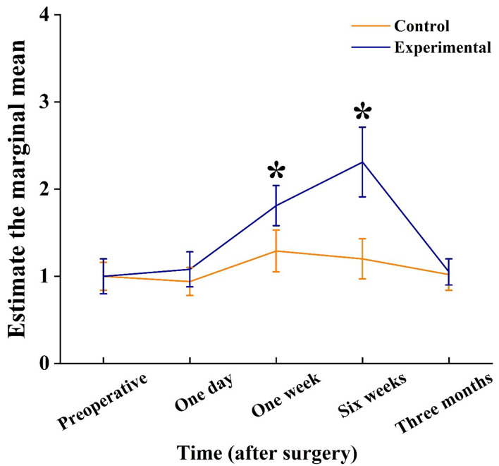
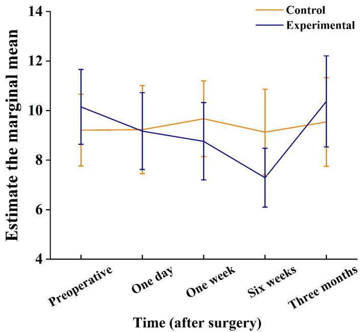
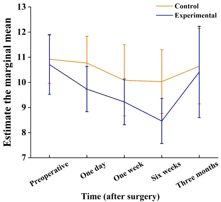
Results: A total of 80 patients (80 eyes) were selected for phacoemulsification treatment with cataract, TF-OSI value of the experimental group was higher than that of the control group at 1 day, 1 week, 6 weeks after surgery (P < 0.05). BUT value was lower than that of the control group at 1 day, 1 week, 6 weeks after surgery (P < 0.05). Schirmer I test value was lower than that of the control group at 1 week and 6 weeks after surgery (P < 0.05). OSDI scores were higher than that of the control group 1 week, 6 weeks and 3 months after surgery (P < 0.05).
Conclusion: Phacoemulsification with LRIs may cause more tissue damage on the ocular surface compared to standard phacoemulsification alone.

 求助内容:
求助内容: 应助结果提醒方式:


