Yu Duan, Kuan Lv, Chao Zhao, Liangbo Han, Jianke Wang, Chuanpeng Zhang, Ziyi Zhang, Hanlin Liu, Ke Yang, Zhen Yuan, Li Zhu, Yuli Wang, Jixin Luan, Guolin Ma, Jiang Liu
{"title":"探索面肌痉挛的面部核中心连通性:对发病机制和手术影响的新见解。","authors":"Yu Duan, Kuan Lv, Chao Zhao, Liangbo Han, Jianke Wang, Chuanpeng Zhang, Ziyi Zhang, Hanlin Liu, Ke Yang, Zhen Yuan, Li Zhu, Yuli Wang, Jixin Luan, Guolin Ma, Jiang Liu","doi":"10.1007/s10548-025-01133-y","DOIUrl":null,"url":null,"abstract":"<p><p>Hemifacial spasm (HFS) is a chronic neurological disorder characterized by involuntary muscle contractions of the face, significantly impacting patients' quality of life. Although the facial nerve nucleus has been implicated in HFS pathogenesis, specific research on its functional connectivity within whole-brain networks remains limited. This study aimed to investigate alterations in whole-brain functional connectivity with the facial nerve nucleus as the region of interest (ROI) in HFS patients, before and after microvascular decompression (MVD), to uncover potential mechanisms underlying the disorder and the impact of surgical intervention. Resting-state functional magnetic resonance imaging (rs-fMRI) was conducted on 30 HFS patients and 30 matched healthy controls. Functional connectivity (FC) was analyzed using the facial nerve nucleus as the seed ROI. Demographic, clinical, and laboratory data were collected, including spasm severity, anxiety and depression scores, and preoperative biomarkers. Statistical analyses assessed differences in FC and its correlation with clinical parameters. HFS patients demonstrated significantly increased FC between the left facial nucleus and the right parahippocampal gyrus, as well as between the right facial nucleus and the right fusiform gyrus, compared to healthy controls. These patterns persisted postoperatively, with additional increased FC observed between the right facial nucleus and bilateral superior temporal gyri. Correlation analyses revealed that left facial nucleus-right parahippocampal gyrus FC was positively associated with spasm severity and fibrinogen levels, while right facial nucleus-right fusiform gyrus FC was negatively correlated with monoamine oxidase (MAO) levels. ReHo of both facial nucleus showed significant differences between preoperative HFS patients and healthy controls, whereas ALFF/fALFF and lateralisation of facial nucleus did not show significant between-group differences. This study highlights the role of altered FC between the facial nucleus and brain regions involved in memory, emotion, and visual processing in HFS pathogenesis. While MVD provides symptomatic relief, its short-term effects on FC appear limited, suggesting that functional connectivity changes are chronic and may serve as biomarkers for disease monitoring. These findings provide novel insights into the neural mechanisms of HFS and emphasize the need for further research on long-term brain network adaptations post-surgery.</p>","PeriodicalId":55329,"journal":{"name":"Brain Topography","volume":"38 5","pages":"58"},"PeriodicalIF":2.9000,"publicationDate":"2025-08-18","publicationTypes":"Journal Article","fieldsOfStudy":null,"isOpenAccess":false,"openAccessPdf":"https://www.ncbi.nlm.nih.gov/pmc/articles/PMC12358327/pdf/","citationCount":"0","resultStr":"{\"title\":\"Exploring Facial Nucleus-Centered Connectivity in Hemifacial Spasm: Novel Insights into Pathogenesis and Surgical Impact.\",\"authors\":\"Yu Duan, Kuan Lv, Chao Zhao, Liangbo Han, Jianke Wang, Chuanpeng Zhang, Ziyi Zhang, Hanlin Liu, Ke Yang, Zhen Yuan, Li Zhu, Yuli Wang, Jixin Luan, Guolin Ma, Jiang Liu\",\"doi\":\"10.1007/s10548-025-01133-y\",\"DOIUrl\":null,\"url\":null,\"abstract\":\"<p><p>Hemifacial spasm (HFS) is a chronic neurological disorder characterized by involuntary muscle contractions of the face, significantly impacting patients' quality of life. Although the facial nerve nucleus has been implicated in HFS pathogenesis, specific research on its functional connectivity within whole-brain networks remains limited. This study aimed to investigate alterations in whole-brain functional connectivity with the facial nerve nucleus as the region of interest (ROI) in HFS patients, before and after microvascular decompression (MVD), to uncover potential mechanisms underlying the disorder and the impact of surgical intervention. Resting-state functional magnetic resonance imaging (rs-fMRI) was conducted on 30 HFS patients and 30 matched healthy controls. Functional connectivity (FC) was analyzed using the facial nerve nucleus as the seed ROI. Demographic, clinical, and laboratory data were collected, including spasm severity, anxiety and depression scores, and preoperative biomarkers. Statistical analyses assessed differences in FC and its correlation with clinical parameters. HFS patients demonstrated significantly increased FC between the left facial nucleus and the right parahippocampal gyrus, as well as between the right facial nucleus and the right fusiform gyrus, compared to healthy controls. These patterns persisted postoperatively, with additional increased FC observed between the right facial nucleus and bilateral superior temporal gyri. Correlation analyses revealed that left facial nucleus-right parahippocampal gyrus FC was positively associated with spasm severity and fibrinogen levels, while right facial nucleus-right fusiform gyrus FC was negatively correlated with monoamine oxidase (MAO) levels. ReHo of both facial nucleus showed significant differences between preoperative HFS patients and healthy controls, whereas ALFF/fALFF and lateralisation of facial nucleus did not show significant between-group differences. This study highlights the role of altered FC between the facial nucleus and brain regions involved in memory, emotion, and visual processing in HFS pathogenesis. While MVD provides symptomatic relief, its short-term effects on FC appear limited, suggesting that functional connectivity changes are chronic and may serve as biomarkers for disease monitoring. These findings provide novel insights into the neural mechanisms of HFS and emphasize the need for further research on long-term brain network adaptations post-surgery.</p>\",\"PeriodicalId\":55329,\"journal\":{\"name\":\"Brain Topography\",\"volume\":\"38 5\",\"pages\":\"58\"},\"PeriodicalIF\":2.9000,\"publicationDate\":\"2025-08-18\",\"publicationTypes\":\"Journal Article\",\"fieldsOfStudy\":null,\"isOpenAccess\":false,\"openAccessPdf\":\"https://www.ncbi.nlm.nih.gov/pmc/articles/PMC12358327/pdf/\",\"citationCount\":\"0\",\"resultStr\":null,\"platform\":\"Semanticscholar\",\"paperid\":null,\"PeriodicalName\":\"Brain Topography\",\"FirstCategoryId\":\"3\",\"ListUrlMain\":\"https://doi.org/10.1007/s10548-025-01133-y\",\"RegionNum\":3,\"RegionCategory\":\"医学\",\"ArticlePicture\":[],\"TitleCN\":null,\"AbstractTextCN\":null,\"PMCID\":null,\"EPubDate\":\"\",\"PubModel\":\"\",\"JCR\":\"Q3\",\"JCRName\":\"CLINICAL NEUROLOGY\",\"Score\":null,\"Total\":0}","platform":"Semanticscholar","paperid":null,"PeriodicalName":"Brain Topography","FirstCategoryId":"3","ListUrlMain":"https://doi.org/10.1007/s10548-025-01133-y","RegionNum":3,"RegionCategory":"医学","ArticlePicture":[],"TitleCN":null,"AbstractTextCN":null,"PMCID":null,"EPubDate":"","PubModel":"","JCR":"Q3","JCRName":"CLINICAL NEUROLOGY","Score":null,"Total":0}
Exploring Facial Nucleus-Centered Connectivity in Hemifacial Spasm: Novel Insights into Pathogenesis and Surgical Impact.
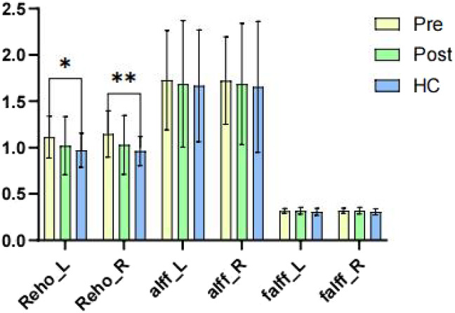
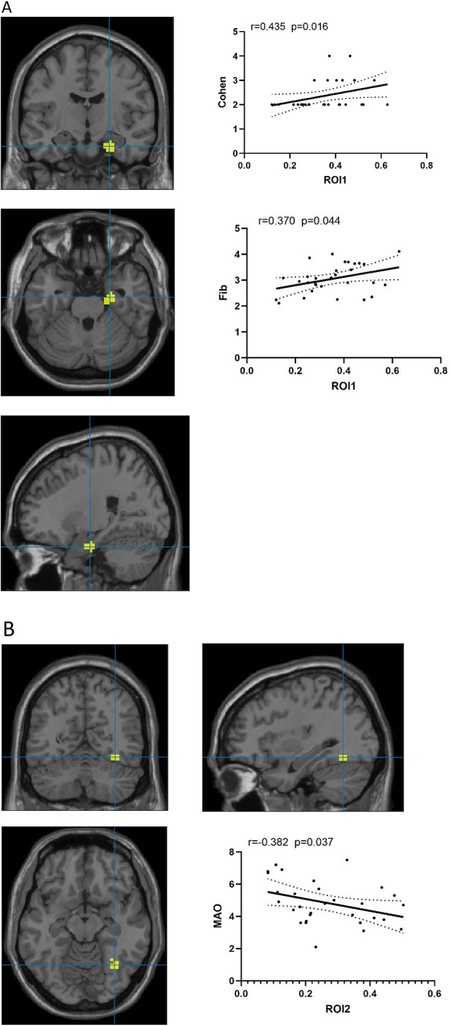
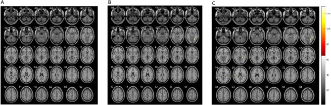
Hemifacial spasm (HFS) is a chronic neurological disorder characterized by involuntary muscle contractions of the face, significantly impacting patients' quality of life. Although the facial nerve nucleus has been implicated in HFS pathogenesis, specific research on its functional connectivity within whole-brain networks remains limited. This study aimed to investigate alterations in whole-brain functional connectivity with the facial nerve nucleus as the region of interest (ROI) in HFS patients, before and after microvascular decompression (MVD), to uncover potential mechanisms underlying the disorder and the impact of surgical intervention. Resting-state functional magnetic resonance imaging (rs-fMRI) was conducted on 30 HFS patients and 30 matched healthy controls. Functional connectivity (FC) was analyzed using the facial nerve nucleus as the seed ROI. Demographic, clinical, and laboratory data were collected, including spasm severity, anxiety and depression scores, and preoperative biomarkers. Statistical analyses assessed differences in FC and its correlation with clinical parameters. HFS patients demonstrated significantly increased FC between the left facial nucleus and the right parahippocampal gyrus, as well as between the right facial nucleus and the right fusiform gyrus, compared to healthy controls. These patterns persisted postoperatively, with additional increased FC observed between the right facial nucleus and bilateral superior temporal gyri. Correlation analyses revealed that left facial nucleus-right parahippocampal gyrus FC was positively associated with spasm severity and fibrinogen levels, while right facial nucleus-right fusiform gyrus FC was negatively correlated with monoamine oxidase (MAO) levels. ReHo of both facial nucleus showed significant differences between preoperative HFS patients and healthy controls, whereas ALFF/fALFF and lateralisation of facial nucleus did not show significant between-group differences. This study highlights the role of altered FC between the facial nucleus and brain regions involved in memory, emotion, and visual processing in HFS pathogenesis. While MVD provides symptomatic relief, its short-term effects on FC appear limited, suggesting that functional connectivity changes are chronic and may serve as biomarkers for disease monitoring. These findings provide novel insights into the neural mechanisms of HFS and emphasize the need for further research on long-term brain network adaptations post-surgery.
期刊介绍:
Brain Topography publishes clinical and basic research on cognitive neuroscience and functional neurophysiology using the full range of imaging techniques including EEG, MEG, fMRI, TMS, diffusion imaging, spectroscopy, intracranial recordings, lesion studies, and related methods. Submissions combining multiple techniques are particularly encouraged, as well as reports of new and innovative methodologies.
 求助内容:
求助内容: 应助结果提醒方式:


