Sarah Abendanan, David Shaul, J Moshe Gomori, Rachel Katz-Brull
{"title":"氘代谢磁共振波谱法研究离体灌注大鼠脑切片缺血再灌注的可行性。","authors":"Sarah Abendanan, David Shaul, J Moshe Gomori, Rachel Katz-Brull","doi":"10.1002/nbm.70115","DOIUrl":null,"url":null,"abstract":"<p><p>Investigating glucose metabolism in the brain using [6,6-<sup>2</sup>H<sub>2</sub>]glucose (<sup>2</sup>H<sub>2</sub>-Glc) and deuterium-based NMR spectroscopy has shown promise for noninvasive monitoring of the fate of this labeled compound. This approach has already been applied in vivo in small animals and human subjects. A model of perfused rat brain slices recently showed promise for the investigation of the metabolic consequences of acute ischemic stroke, which is a significant cause of death and morbidity worldwide. The current study aimed to implement the deuterium-based glucose metabolism monitoring approach to study the metabolic consequences of ischemia and reperfusion in the rat brain ex vivo. In agreement with previous studies, we found that deuterated lactate (<sup>2</sup>H<sub>2</sub>-Lac) was immediately formed in the brain upon administration of <sup>2</sup>H<sub>2</sub>-Glc to the perfusion medium. This metabolite remained the predominant metabolic fate observed in the <sup>2</sup>H-NMR spectra. Upon perfusion arrest, <sup>2</sup>H<sub>2</sub>-Lac quickly built up to the same amount of <sup>2</sup>H<sub>2</sub>-Glc eliminated from the medium engulfing the slices, reaching fivefold to sixfold its baseline level (n = 6, three animals, and two ischemic conditions in each). Upon reperfusion, <sup>2</sup>H<sub>2</sub>-Lac decreased to its level before the ischemic condition, and <sup>2</sup>H<sub>2</sub>-Glc returned to its baseline. <sup>2</sup>H<sub>2</sub>-Lac washout to the medium amounted to 2.2% of the <sup>2</sup>H<sub>2</sub>-Lac signal associated with the slices after about 5 h of perfusion with <sup>2</sup>H<sub>2</sub>-Glc, suggesting that the <sup>2</sup>H<sub>2</sub>-Lac signal observed during the experiments was predominantly intracellular. These results demonstrate the utility of <sup>2</sup>H<sub>2</sub>-Glc and <sup>2</sup>H-NMR in monitoring the consequences of ischemia and reperfusion in the perfused rat brain slices model.</p>","PeriodicalId":19309,"journal":{"name":"NMR in Biomedicine","volume":"38 9","pages":"e70115"},"PeriodicalIF":2.7000,"publicationDate":"2025-09-01","publicationTypes":"Journal Article","fieldsOfStudy":null,"isOpenAccess":false,"openAccessPdf":"https://www.ncbi.nlm.nih.gov/pmc/articles/PMC12358337/pdf/","citationCount":"0","resultStr":"{\"title\":\"Feasibility of Deuterium Metabolic Magnetic Resonance Spectroscopy for the Investigation of Ischemia and Reperfusion in Rat Brain Slices Perfused Ex Vivo.\",\"authors\":\"Sarah Abendanan, David Shaul, J Moshe Gomori, Rachel Katz-Brull\",\"doi\":\"10.1002/nbm.70115\",\"DOIUrl\":null,\"url\":null,\"abstract\":\"<p><p>Investigating glucose metabolism in the brain using [6,6-<sup>2</sup>H<sub>2</sub>]glucose (<sup>2</sup>H<sub>2</sub>-Glc) and deuterium-based NMR spectroscopy has shown promise for noninvasive monitoring of the fate of this labeled compound. This approach has already been applied in vivo in small animals and human subjects. A model of perfused rat brain slices recently showed promise for the investigation of the metabolic consequences of acute ischemic stroke, which is a significant cause of death and morbidity worldwide. The current study aimed to implement the deuterium-based glucose metabolism monitoring approach to study the metabolic consequences of ischemia and reperfusion in the rat brain ex vivo. In agreement with previous studies, we found that deuterated lactate (<sup>2</sup>H<sub>2</sub>-Lac) was immediately formed in the brain upon administration of <sup>2</sup>H<sub>2</sub>-Glc to the perfusion medium. This metabolite remained the predominant metabolic fate observed in the <sup>2</sup>H-NMR spectra. Upon perfusion arrest, <sup>2</sup>H<sub>2</sub>-Lac quickly built up to the same amount of <sup>2</sup>H<sub>2</sub>-Glc eliminated from the medium engulfing the slices, reaching fivefold to sixfold its baseline level (n = 6, three animals, and two ischemic conditions in each). Upon reperfusion, <sup>2</sup>H<sub>2</sub>-Lac decreased to its level before the ischemic condition, and <sup>2</sup>H<sub>2</sub>-Glc returned to its baseline. <sup>2</sup>H<sub>2</sub>-Lac washout to the medium amounted to 2.2% of the <sup>2</sup>H<sub>2</sub>-Lac signal associated with the slices after about 5 h of perfusion with <sup>2</sup>H<sub>2</sub>-Glc, suggesting that the <sup>2</sup>H<sub>2</sub>-Lac signal observed during the experiments was predominantly intracellular. These results demonstrate the utility of <sup>2</sup>H<sub>2</sub>-Glc and <sup>2</sup>H-NMR in monitoring the consequences of ischemia and reperfusion in the perfused rat brain slices model.</p>\",\"PeriodicalId\":19309,\"journal\":{\"name\":\"NMR in Biomedicine\",\"volume\":\"38 9\",\"pages\":\"e70115\"},\"PeriodicalIF\":2.7000,\"publicationDate\":\"2025-09-01\",\"publicationTypes\":\"Journal Article\",\"fieldsOfStudy\":null,\"isOpenAccess\":false,\"openAccessPdf\":\"https://www.ncbi.nlm.nih.gov/pmc/articles/PMC12358337/pdf/\",\"citationCount\":\"0\",\"resultStr\":null,\"platform\":\"Semanticscholar\",\"paperid\":null,\"PeriodicalName\":\"NMR in Biomedicine\",\"FirstCategoryId\":\"3\",\"ListUrlMain\":\"https://doi.org/10.1002/nbm.70115\",\"RegionNum\":4,\"RegionCategory\":\"医学\",\"ArticlePicture\":[],\"TitleCN\":null,\"AbstractTextCN\":null,\"PMCID\":null,\"EPubDate\":\"\",\"PubModel\":\"\",\"JCR\":\"Q2\",\"JCRName\":\"BIOPHYSICS\",\"Score\":null,\"Total\":0}","platform":"Semanticscholar","paperid":null,"PeriodicalName":"NMR in Biomedicine","FirstCategoryId":"3","ListUrlMain":"https://doi.org/10.1002/nbm.70115","RegionNum":4,"RegionCategory":"医学","ArticlePicture":[],"TitleCN":null,"AbstractTextCN":null,"PMCID":null,"EPubDate":"","PubModel":"","JCR":"Q2","JCRName":"BIOPHYSICS","Score":null,"Total":0}
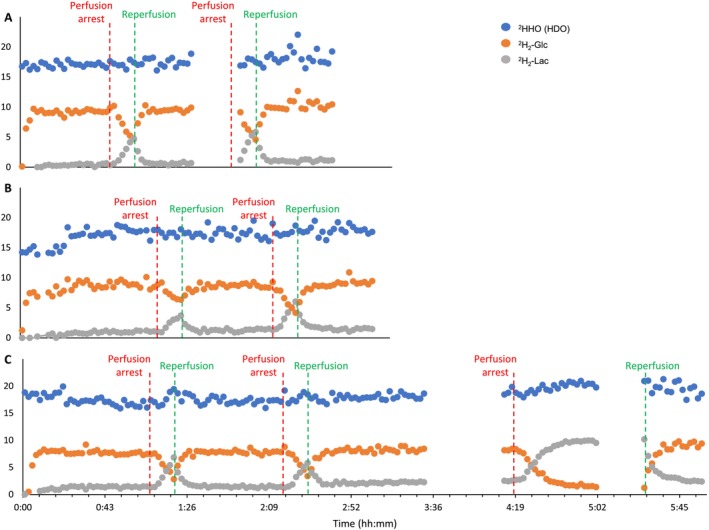
Feasibility of Deuterium Metabolic Magnetic Resonance Spectroscopy for the Investigation of Ischemia and Reperfusion in Rat Brain Slices Perfused Ex Vivo.
Investigating glucose metabolism in the brain using [6,6-2H2]glucose (2H2-Glc) and deuterium-based NMR spectroscopy has shown promise for noninvasive monitoring of the fate of this labeled compound. This approach has already been applied in vivo in small animals and human subjects. A model of perfused rat brain slices recently showed promise for the investigation of the metabolic consequences of acute ischemic stroke, which is a significant cause of death and morbidity worldwide. The current study aimed to implement the deuterium-based glucose metabolism monitoring approach to study the metabolic consequences of ischemia and reperfusion in the rat brain ex vivo. In agreement with previous studies, we found that deuterated lactate (2H2-Lac) was immediately formed in the brain upon administration of 2H2-Glc to the perfusion medium. This metabolite remained the predominant metabolic fate observed in the 2H-NMR spectra. Upon perfusion arrest, 2H2-Lac quickly built up to the same amount of 2H2-Glc eliminated from the medium engulfing the slices, reaching fivefold to sixfold its baseline level (n = 6, three animals, and two ischemic conditions in each). Upon reperfusion, 2H2-Lac decreased to its level before the ischemic condition, and 2H2-Glc returned to its baseline. 2H2-Lac washout to the medium amounted to 2.2% of the 2H2-Lac signal associated with the slices after about 5 h of perfusion with 2H2-Glc, suggesting that the 2H2-Lac signal observed during the experiments was predominantly intracellular. These results demonstrate the utility of 2H2-Glc and 2H-NMR in monitoring the consequences of ischemia and reperfusion in the perfused rat brain slices model.
期刊介绍:
NMR in Biomedicine is a journal devoted to the publication of original full-length papers, rapid communications and review articles describing the development of magnetic resonance spectroscopy or imaging methods or their use to investigate physiological, biochemical, biophysical or medical problems. Topics for submitted papers should be in one of the following general categories: (a) development of methods and instrumentation for MR of biological systems; (b) studies of normal or diseased organs, tissues or cells; (c) diagnosis or treatment of disease. Reports may cover work on patients or healthy human subjects, in vivo animal experiments, studies of isolated organs or cultured cells, analysis of tissue extracts, NMR theory, experimental techniques, or instrumentation.


 求助内容:
求助内容: 应助结果提醒方式:


