Ramy Abdul-Rahman Ishaq, Maged S Alhammadi, Mona M Salah Fayed, Sherif A Elkordy, Najah Alhashimi, Abeer A Almashraqi
{"title":"使用forsus抗疲劳装置治疗的患者的位置和尺寸颞下颌关节骨改变:一项非随机临床试验。","authors":"Ramy Abdul-Rahman Ishaq, Maged S Alhammadi, Mona M Salah Fayed, Sherif A Elkordy, Najah Alhashimi, Abeer A Almashraqi","doi":"10.1007/s00784-025-06474-3","DOIUrl":null,"url":null,"abstract":"<p><strong>Objective: </strong>This study aimed to assess the osseous positional and dimensional changes in the temporomandibular joint (TMJ) of patients with skeletal Class II malocclusion treated with the Forsus Fatigue Resistant Device (FFRD).</p><p><strong>Materials and methods: </strong>This non-randomized clinical trial included 40 female subjects, aged 11 to 15, with skeletal Class II malocclusion. Participants were divided into a treatment and a control group. After alignment and leveling with fixed orthodontic appliances using 0.019 × 0.025-inch stainless-steel archwires, the FFRD was fitted. The overjet was corrected to achieve an edge-to-edge incisor relationship. Cone Beam Computed Tomography (CBCT) images were taken before (T1) and after (T2) the fixed functional phase. The TMJs were assessed for positional and dimensional osseous changes in the mandibular condyles, glenoid fossae, and joint spaces. Intra- and inter-group comparisons were conducted using paired t-tests and independent t-tests, respectively.</p><p><strong>Results: </strong>The initial measurements of age, cervical stage, anteroposterior and vertical skeletal alignment, and TMJ parameters were similar between the study and control groups. Three participants from the study group were lost to follow-up, resulting in 17 participants completing the trial. In the treatment group, condylar width decreased significantly by 0.52 ± 0.92 mm, in contrast to an increase of 0.17 ± 0.35 mm in the control group (P = 0.010). Moreover, the anterior wall inclination in the treatment group was reduced by 3.13 ± 10.77 degrees, compared to an increase of 2.95 ± 4.1 degrees in the control group (P = 0.003). All other measurements displayed no significant differences between the two groups.</p><p><strong>Conclusion: </strong>In the short term, the FFRD redirected the growth of the articular eminence anteriorly, contrasting with the normal growth pattern of untreated individuals. However, no additional positional or dimensional changes in the TMJ were observed.</p><p><strong>Clinical relevance: </strong>By aligning the jaw and correcting overjet, clinicians can potentially enhance occlusal relationships and contribute to better jaw function. However, it is important to investigate whether this process is associated with any changes in the bony structures of the TMJ. This study underscores the efficacy of the FFRD in reshaping the osseous components of the TMJ, which may lead to improved functional outcomes for patients with skeletal Class II malocclusion.</p>","PeriodicalId":10461,"journal":{"name":"Clinical Oral Investigations","volume":"29 9","pages":"414"},"PeriodicalIF":3.1000,"publicationDate":"2025-08-18","publicationTypes":"Journal Article","fieldsOfStudy":null,"isOpenAccess":false,"openAccessPdf":"https://www.ncbi.nlm.nih.gov/pmc/articles/PMC12358331/pdf/","citationCount":"0","resultStr":"{\"title\":\"Positional and dimensional temporomandibular joint osseous changes in patients treated with the forsus fatigue resistant device: a non-randomized clinical trial.\",\"authors\":\"Ramy Abdul-Rahman Ishaq, Maged S Alhammadi, Mona M Salah Fayed, Sherif A Elkordy, Najah Alhashimi, Abeer A Almashraqi\",\"doi\":\"10.1007/s00784-025-06474-3\",\"DOIUrl\":null,\"url\":null,\"abstract\":\"<p><strong>Objective: </strong>This study aimed to assess the osseous positional and dimensional changes in the temporomandibular joint (TMJ) of patients with skeletal Class II malocclusion treated with the Forsus Fatigue Resistant Device (FFRD).</p><p><strong>Materials and methods: </strong>This non-randomized clinical trial included 40 female subjects, aged 11 to 15, with skeletal Class II malocclusion. Participants were divided into a treatment and a control group. After alignment and leveling with fixed orthodontic appliances using 0.019 × 0.025-inch stainless-steel archwires, the FFRD was fitted. The overjet was corrected to achieve an edge-to-edge incisor relationship. Cone Beam Computed Tomography (CBCT) images were taken before (T1) and after (T2) the fixed functional phase. The TMJs were assessed for positional and dimensional osseous changes in the mandibular condyles, glenoid fossae, and joint spaces. Intra- and inter-group comparisons were conducted using paired t-tests and independent t-tests, respectively.</p><p><strong>Results: </strong>The initial measurements of age, cervical stage, anteroposterior and vertical skeletal alignment, and TMJ parameters were similar between the study and control groups. Three participants from the study group were lost to follow-up, resulting in 17 participants completing the trial. In the treatment group, condylar width decreased significantly by 0.52 ± 0.92 mm, in contrast to an increase of 0.17 ± 0.35 mm in the control group (P = 0.010). Moreover, the anterior wall inclination in the treatment group was reduced by 3.13 ± 10.77 degrees, compared to an increase of 2.95 ± 4.1 degrees in the control group (P = 0.003). All other measurements displayed no significant differences between the two groups.</p><p><strong>Conclusion: </strong>In the short term, the FFRD redirected the growth of the articular eminence anteriorly, contrasting with the normal growth pattern of untreated individuals. However, no additional positional or dimensional changes in the TMJ were observed.</p><p><strong>Clinical relevance: </strong>By aligning the jaw and correcting overjet, clinicians can potentially enhance occlusal relationships and contribute to better jaw function. However, it is important to investigate whether this process is associated with any changes in the bony structures of the TMJ. This study underscores the efficacy of the FFRD in reshaping the osseous components of the TMJ, which may lead to improved functional outcomes for patients with skeletal Class II malocclusion.</p>\",\"PeriodicalId\":10461,\"journal\":{\"name\":\"Clinical Oral Investigations\",\"volume\":\"29 9\",\"pages\":\"414\"},\"PeriodicalIF\":3.1000,\"publicationDate\":\"2025-08-18\",\"publicationTypes\":\"Journal Article\",\"fieldsOfStudy\":null,\"isOpenAccess\":false,\"openAccessPdf\":\"https://www.ncbi.nlm.nih.gov/pmc/articles/PMC12358331/pdf/\",\"citationCount\":\"0\",\"resultStr\":null,\"platform\":\"Semanticscholar\",\"paperid\":null,\"PeriodicalName\":\"Clinical Oral Investigations\",\"FirstCategoryId\":\"3\",\"ListUrlMain\":\"https://doi.org/10.1007/s00784-025-06474-3\",\"RegionNum\":2,\"RegionCategory\":\"医学\",\"ArticlePicture\":[],\"TitleCN\":null,\"AbstractTextCN\":null,\"PMCID\":null,\"EPubDate\":\"\",\"PubModel\":\"\",\"JCR\":\"Q1\",\"JCRName\":\"DENTISTRY, ORAL SURGERY & MEDICINE\",\"Score\":null,\"Total\":0}","platform":"Semanticscholar","paperid":null,"PeriodicalName":"Clinical Oral Investigations","FirstCategoryId":"3","ListUrlMain":"https://doi.org/10.1007/s00784-025-06474-3","RegionNum":2,"RegionCategory":"医学","ArticlePicture":[],"TitleCN":null,"AbstractTextCN":null,"PMCID":null,"EPubDate":"","PubModel":"","JCR":"Q1","JCRName":"DENTISTRY, ORAL SURGERY & MEDICINE","Score":null,"Total":0}
Positional and dimensional temporomandibular joint osseous changes in patients treated with the forsus fatigue resistant device: a non-randomized clinical trial.
Objective: This study aimed to assess the osseous positional and dimensional changes in the temporomandibular joint (TMJ) of patients with skeletal Class II malocclusion treated with the Forsus Fatigue Resistant Device (FFRD).
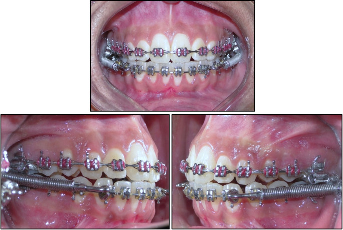
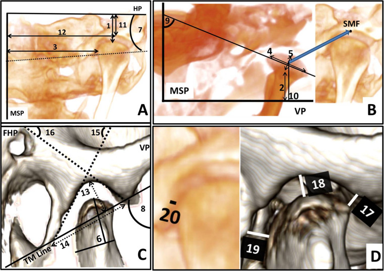
Materials and methods: This non-randomized clinical trial included 40 female subjects, aged 11 to 15, with skeletal Class II malocclusion. Participants were divided into a treatment and a control group. After alignment and leveling with fixed orthodontic appliances using 0.019 × 0.025-inch stainless-steel archwires, the FFRD was fitted. The overjet was corrected to achieve an edge-to-edge incisor relationship. Cone Beam Computed Tomography (CBCT) images were taken before (T1) and after (T2) the fixed functional phase. The TMJs were assessed for positional and dimensional osseous changes in the mandibular condyles, glenoid fossae, and joint spaces. Intra- and inter-group comparisons were conducted using paired t-tests and independent t-tests, respectively.
Results: The initial measurements of age, cervical stage, anteroposterior and vertical skeletal alignment, and TMJ parameters were similar between the study and control groups. Three participants from the study group were lost to follow-up, resulting in 17 participants completing the trial. In the treatment group, condylar width decreased significantly by 0.52 ± 0.92 mm, in contrast to an increase of 0.17 ± 0.35 mm in the control group (P = 0.010). Moreover, the anterior wall inclination in the treatment group was reduced by 3.13 ± 10.77 degrees, compared to an increase of 2.95 ± 4.1 degrees in the control group (P = 0.003). All other measurements displayed no significant differences between the two groups.
Conclusion: In the short term, the FFRD redirected the growth of the articular eminence anteriorly, contrasting with the normal growth pattern of untreated individuals. However, no additional positional or dimensional changes in the TMJ were observed.
Clinical relevance: By aligning the jaw and correcting overjet, clinicians can potentially enhance occlusal relationships and contribute to better jaw function. However, it is important to investigate whether this process is associated with any changes in the bony structures of the TMJ. This study underscores the efficacy of the FFRD in reshaping the osseous components of the TMJ, which may lead to improved functional outcomes for patients with skeletal Class II malocclusion.
期刊介绍:
The journal Clinical Oral Investigations is a multidisciplinary, international forum for publication of research from all fields of oral medicine. The journal publishes original scientific articles and invited reviews which provide up-to-date results of basic and clinical studies in oral and maxillofacial science and medicine. The aim is to clarify the relevance of new results to modern practice, for an international readership. Coverage includes maxillofacial and oral surgery, prosthetics and restorative dentistry, operative dentistry, endodontics, periodontology, orthodontics, dental materials science, clinical trials, epidemiology, pedodontics, oral implant, preventive dentistiry, oral pathology, oral basic sciences and more.

 求助内容:
求助内容: 应助结果提醒方式:


