Rachel E. Rubino, Martin Kaufmann, Amoon Jamzad, Natasha Iaboni, Kevin Yi Mi Ren, Jian Yu, Haidy Metwally, Manuela Kunz, John F. Rudan, Parvin Mousavi, Gabor Fichtinger, Richard Oleschuk, Christopher J. B. Nicol
{"title":"利用3D体外实验评估人类乳腺癌细胞的快速蒸发电离质谱分析。","authors":"Rachel E. Rubino, Martin Kaufmann, Amoon Jamzad, Natasha Iaboni, Kevin Yi Mi Ren, Jian Yu, Haidy Metwally, Manuela Kunz, John F. Rudan, Parvin Mousavi, Gabor Fichtinger, Richard Oleschuk, Christopher J. B. Nicol","doi":"10.1007/s00216-025-06035-3","DOIUrl":null,"url":null,"abstract":"<div><p>Rapid evaporative ionization mass spectrometry (REIMS) enables near real-time sampling and classification of tissues on the basis of lipid and small molecule profiles. Because samples are ablated during REIMS, validation of mass spectra by assignment of class labels using gold-standard methods remains challenging. Further, determining the number of abnormal cells within a mixture of normal cells by REIMS remains an elusive but critical parameter when evaluating the utility of REIMS for use in clinical settings. To address this, we developed a three-dimensional assay based on fluorescently labelled human breast cell lines, MCF-10A (pseudo-normal) and MDA-MB-231 (malignant), embedded in matrigel cubes that enables calculation of cell numbers ablated by REIMS by measuring fluorescence before and after REIMS sampling. We observed a reasonably uniform distribution of cells throughout the matrigel matrix, which yielded REIMS spectra rich in complex glycerophospholipids resembling solid tissues and cell pellets. A non-linear relationship between signal intensity and the number of cells ablated was observed. We trained multivariate models using 10% increments of MDA-MB-231 cells mixed with MCF-10A, and blindly tested the models on separate cubes containing 1–100% MDA-MB-231s. Percent difference from target MDA-MB-231 cell composition was within 34% for cubes containing as little as 5% MDA-MB-231 cells. Overall, our study highlights how a simple 3D in vitro approach may help reveal the quantitative potential of mass spectral profiling methods such as REIMS to assist with validating REIMS spectra containing mixtures of different cell lines. This assay platform may help calibrate REIMS-based methods to recognize specific cell type mixtures encountered at surgical resection margins.\n</p></div>","PeriodicalId":462,"journal":{"name":"Analytical and Bioanalytical Chemistry","volume":"417 22","pages":"5115 - 5130"},"PeriodicalIF":3.8000,"publicationDate":"2025-08-18","publicationTypes":"Journal Article","fieldsOfStudy":null,"isOpenAccess":false,"openAccessPdf":"","citationCount":"0","resultStr":"{\"title\":\"Evaluating rapid evaporative ionization mass spectrometry profiles of human breast cancer cells using a 3D in vitro assay\",\"authors\":\"Rachel E. Rubino, Martin Kaufmann, Amoon Jamzad, Natasha Iaboni, Kevin Yi Mi Ren, Jian Yu, Haidy Metwally, Manuela Kunz, John F. Rudan, Parvin Mousavi, Gabor Fichtinger, Richard Oleschuk, Christopher J. B. Nicol\",\"doi\":\"10.1007/s00216-025-06035-3\",\"DOIUrl\":null,\"url\":null,\"abstract\":\"<div><p>Rapid evaporative ionization mass spectrometry (REIMS) enables near real-time sampling and classification of tissues on the basis of lipid and small molecule profiles. Because samples are ablated during REIMS, validation of mass spectra by assignment of class labels using gold-standard methods remains challenging. Further, determining the number of abnormal cells within a mixture of normal cells by REIMS remains an elusive but critical parameter when evaluating the utility of REIMS for use in clinical settings. To address this, we developed a three-dimensional assay based on fluorescently labelled human breast cell lines, MCF-10A (pseudo-normal) and MDA-MB-231 (malignant), embedded in matrigel cubes that enables calculation of cell numbers ablated by REIMS by measuring fluorescence before and after REIMS sampling. We observed a reasonably uniform distribution of cells throughout the matrigel matrix, which yielded REIMS spectra rich in complex glycerophospholipids resembling solid tissues and cell pellets. A non-linear relationship between signal intensity and the number of cells ablated was observed. We trained multivariate models using 10% increments of MDA-MB-231 cells mixed with MCF-10A, and blindly tested the models on separate cubes containing 1–100% MDA-MB-231s. Percent difference from target MDA-MB-231 cell composition was within 34% for cubes containing as little as 5% MDA-MB-231 cells. Overall, our study highlights how a simple 3D in vitro approach may help reveal the quantitative potential of mass spectral profiling methods such as REIMS to assist with validating REIMS spectra containing mixtures of different cell lines. This assay platform may help calibrate REIMS-based methods to recognize specific cell type mixtures encountered at surgical resection margins.\\n</p></div>\",\"PeriodicalId\":462,\"journal\":{\"name\":\"Analytical and Bioanalytical Chemistry\",\"volume\":\"417 22\",\"pages\":\"5115 - 5130\"},\"PeriodicalIF\":3.8000,\"publicationDate\":\"2025-08-18\",\"publicationTypes\":\"Journal Article\",\"fieldsOfStudy\":null,\"isOpenAccess\":false,\"openAccessPdf\":\"\",\"citationCount\":\"0\",\"resultStr\":null,\"platform\":\"Semanticscholar\",\"paperid\":null,\"PeriodicalName\":\"Analytical and Bioanalytical Chemistry\",\"FirstCategoryId\":\"92\",\"ListUrlMain\":\"https://link.springer.com/article/10.1007/s00216-025-06035-3\",\"RegionNum\":2,\"RegionCategory\":\"化学\",\"ArticlePicture\":[],\"TitleCN\":null,\"AbstractTextCN\":null,\"PMCID\":null,\"EPubDate\":\"\",\"PubModel\":\"\",\"JCR\":\"Q1\",\"JCRName\":\"BIOCHEMICAL RESEARCH METHODS\",\"Score\":null,\"Total\":0}","platform":"Semanticscholar","paperid":null,"PeriodicalName":"Analytical and Bioanalytical Chemistry","FirstCategoryId":"92","ListUrlMain":"https://link.springer.com/article/10.1007/s00216-025-06035-3","RegionNum":2,"RegionCategory":"化学","ArticlePicture":[],"TitleCN":null,"AbstractTextCN":null,"PMCID":null,"EPubDate":"","PubModel":"","JCR":"Q1","JCRName":"BIOCHEMICAL RESEARCH METHODS","Score":null,"Total":0}
Evaluating rapid evaporative ionization mass spectrometry profiles of human breast cancer cells using a 3D in vitro assay
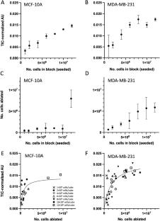
Rapid evaporative ionization mass spectrometry (REIMS) enables near real-time sampling and classification of tissues on the basis of lipid and small molecule profiles. Because samples are ablated during REIMS, validation of mass spectra by assignment of class labels using gold-standard methods remains challenging. Further, determining the number of abnormal cells within a mixture of normal cells by REIMS remains an elusive but critical parameter when evaluating the utility of REIMS for use in clinical settings. To address this, we developed a three-dimensional assay based on fluorescently labelled human breast cell lines, MCF-10A (pseudo-normal) and MDA-MB-231 (malignant), embedded in matrigel cubes that enables calculation of cell numbers ablated by REIMS by measuring fluorescence before and after REIMS sampling. We observed a reasonably uniform distribution of cells throughout the matrigel matrix, which yielded REIMS spectra rich in complex glycerophospholipids resembling solid tissues and cell pellets. A non-linear relationship between signal intensity and the number of cells ablated was observed. We trained multivariate models using 10% increments of MDA-MB-231 cells mixed with MCF-10A, and blindly tested the models on separate cubes containing 1–100% MDA-MB-231s. Percent difference from target MDA-MB-231 cell composition was within 34% for cubes containing as little as 5% MDA-MB-231 cells. Overall, our study highlights how a simple 3D in vitro approach may help reveal the quantitative potential of mass spectral profiling methods such as REIMS to assist with validating REIMS spectra containing mixtures of different cell lines. This assay platform may help calibrate REIMS-based methods to recognize specific cell type mixtures encountered at surgical resection margins.
期刊介绍:
Analytical and Bioanalytical Chemistry’s mission is the rapid publication of excellent and high-impact research articles on fundamental and applied topics of analytical and bioanalytical measurement science. Its scope is broad, and ranges from novel measurement platforms and their characterization to multidisciplinary approaches that effectively address important scientific problems. The Editors encourage submissions presenting innovative analytical research in concept, instrumentation, methods, and/or applications, including: mass spectrometry, spectroscopy, and electroanalysis; advanced separations; analytical strategies in “-omics” and imaging, bioanalysis, and sampling; miniaturized devices, medical diagnostics, sensors; analytical characterization of nano- and biomaterials; chemometrics and advanced data analysis.
 求助内容:
求助内容: 应助结果提醒方式:


