乙醇消耗和曼氏血吸虫感染对小鼠肾脏组织病理学的协同影响。
IF 2.5
3区 医学
Q2 PARASITOLOGY
引用次数: 0
摘要
乙醇摄入合并急性曼氏血吸虫感染对肾脏结构的影响尚不清楚。本研究评估了暴露于这两种条件下的小鼠的肾脏组织病理学。雄性瑞士韦伯斯特小鼠首先感染曼氏梭菌,随后进行一段时间的乙醇或水灌胃。在此暴露期后,将动物分为四个实验组:C(对照组)、UE(未感染 + 乙醇)、I(感染)和IE(感染 + 乙醇)。感染组皮下接种80只曼氏血吸虫尾蚴(BH株)。从感染后第35天开始,乙醇组动物每天灌胃18%乙醇(200 μL),连续28 d。第64天,老鼠被安乐死,它们的肾脏…收集用于组织学分析。组织病理学检查显示,UE组蛋白变性,管腔变窄,近端小管微绒毛丢失,白细胞浸润。I组表现为小管坏死,远端小管有白细胞浸润。IE组表现为蛋白变性、白细胞浸润、系膜细胞增生、肾小球内缩。UE、I、IE组均观察到髓质水肿和白细胞浸润。形态计量学分析显示,与c组相比,UE组(+47%)、I组(+71%)和IE组(+54%)肾小球面积增加,UE组(+42%)、I组(+82%)和IE组(+72%)肾小球丛面积更大。Bowman的空间在UE(+58%)、I(+51%)和IE(+68%)中有所增加。肾小球簇直径在UE(+21%)、I(+39%)和IE(+42%)组增加,IE组比UE组增加17%。与c组相比,UE组(+381%)和IE组(+389%)肾实质胶原沉积明显增加,I组和IE组近端小管体积密度下降22%。远端小管密度在UE (-62%), I(-58%)和IE(-79%)中下降。UE(+14%)、I(+13%)和IE(+16%)的白细胞浸润增加。这些发现表明,乙醇加重了血吸虫病患者的肾脏损害,加剧了炎症和结构损伤。本文章由计算机程序翻译,如有差异,请以英文原文为准。
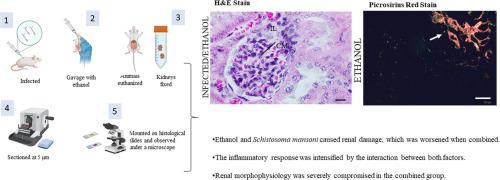
The synergistic impact of ethanol consumption and Schistosoma mansoni infection on renal histopathology in mice
The impact of ethanol ingestion combined with acute Schistosoma mansoni infection on renal architecture remains unclear. This study evaluated renal histopathology in mice exposed to both conditions. Male Swiss Webster mice were first infected with S. mansoni and subsequently subjected to a period of ethanol or water gavage. After this exposure period, animals were allocated into four experimental groups: C (control), UE (uninfected + ethanol), I (infected), and IE (infected + ethanol). The infected groups were subcutaneously inoculated with 80 cercariae of S. mansoni (BH strain). Starting on day 35 post-infection, animals in the ethanol groups received a daily dose of 18 % ethanol by gavage (200 μL) for 28 days. On day 64, the mice were euthanized, and their kidneys were... collected for histological analysis. Histopathological examination revealed that the UE group showed protein denaturation, narrowed tubular lumen, microvilli loss in proximal tubules, and leukocyte infiltration. The I group showed tubular necrosis and leukocyte infiltration in distal tubules. The IE group presented protein denaturation, leukocyte infiltration, mesangial cell proliferation, and glomerular retraction. Medullary edema and leukocyte infiltration were observed in groups UE, I, and IE. Morphometric analysis showed increased glomerular area in groups UE (+47 %), I (+71 %), and IE (+54 %) compared to C. The glomerular tuft area was larger in UE (+42 %), I (+82 %), and IE (+72 %). Bowman's space increased in UE (+58 %), I (+51 %), and IE (+68 %). Glomerular tuft diameter rose in UE (+21 %), I (+39 %), and IE (+42 %), with IE showing a 17 % increase compared to UE. Collagen deposition in renal parenchyma increased markedly in UE (+381 %) and IE (+389 %) versus C. The volume density of proximal tubules decreased 22 % in I and IE. The distal tubule density dropped in UE (-62 %), I (-58 %), and IE (-79 %). Leukocyte infiltration increased in UE (+14 %), I (+13 %), and IE (+16 %). These findings demonstrate that ethanol exacerbates renal damage in schistosomiasis, intensifying inflammation, and structural impairment.
求助全文
通过发布文献求助,成功后即可免费获取论文全文。
去求助
来源期刊
Acta tropica
医学-寄生虫学
CiteScore
5.40
自引率
11.10%
发文量
383
审稿时长
37 days
期刊介绍:
Acta Tropica, is an international journal on infectious diseases that covers public health sciences and biomedical research with particular emphasis on topics relevant to human and animal health in the tropics and the subtropics.
 求助内容:
求助内容: 应助结果提醒方式:


