Behzad Khademi, Mehdi Moallem, Mahsa Kohandel-Shirazi
{"title":"儿童泪囊异位骨化1例。","authors":"Behzad Khademi, Mehdi Moallem, Mahsa Kohandel-Shirazi","doi":"10.18502/jovr.v20.16581","DOIUrl":null,"url":null,"abstract":"<p><strong>Purpose: </strong>We present a case of hemolacria, which emerged as an unusual mass in the lacrimal sac.</p><p><strong>Case report: </strong>A nine-year-old girl presented with intermittent unilateral hemolacria and episodes of dacryocystitis with no further remarkable medical or surgical history. CT scan indicated the presence of well-defined calcified tissue enclosed within the lacrimal sac. Following external dacryocystorhinostomy, well-formed firm tissue resembling bony tissue was extracted and sent for pathological evaluation, along with biopsies from the lacrimal sac. Histopathological analysis revealed heterotopic bone formation with nonspecific inflammation of the lacrimal sac. No underlying cause was discernible in the complementary assessment, and no recurrence was noted at one-year follow-up.</p><p><strong>Conclusion: </strong>A child with hemolacria was found to have heterotopic ossification in the lacrimal sac with no discernible underlying cause.</p>","PeriodicalId":16586,"journal":{"name":"Journal of Ophthalmic & Vision Research","volume":"20 ","pages":""},"PeriodicalIF":1.5000,"publicationDate":"2025-07-30","publicationTypes":"Journal Article","fieldsOfStudy":null,"isOpenAccess":false,"openAccessPdf":"https://www.ncbi.nlm.nih.gov/pmc/articles/PMC12322506/pdf/","citationCount":"0","resultStr":"{\"title\":\"Heterotopic Ossification Originating from the Lacrimal Sac of a Child: A Case Report.\",\"authors\":\"Behzad Khademi, Mehdi Moallem, Mahsa Kohandel-Shirazi\",\"doi\":\"10.18502/jovr.v20.16581\",\"DOIUrl\":null,\"url\":null,\"abstract\":\"<p><strong>Purpose: </strong>We present a case of hemolacria, which emerged as an unusual mass in the lacrimal sac.</p><p><strong>Case report: </strong>A nine-year-old girl presented with intermittent unilateral hemolacria and episodes of dacryocystitis with no further remarkable medical or surgical history. CT scan indicated the presence of well-defined calcified tissue enclosed within the lacrimal sac. Following external dacryocystorhinostomy, well-formed firm tissue resembling bony tissue was extracted and sent for pathological evaluation, along with biopsies from the lacrimal sac. Histopathological analysis revealed heterotopic bone formation with nonspecific inflammation of the lacrimal sac. No underlying cause was discernible in the complementary assessment, and no recurrence was noted at one-year follow-up.</p><p><strong>Conclusion: </strong>A child with hemolacria was found to have heterotopic ossification in the lacrimal sac with no discernible underlying cause.</p>\",\"PeriodicalId\":16586,\"journal\":{\"name\":\"Journal of Ophthalmic & Vision Research\",\"volume\":\"20 \",\"pages\":\"\"},\"PeriodicalIF\":1.5000,\"publicationDate\":\"2025-07-30\",\"publicationTypes\":\"Journal Article\",\"fieldsOfStudy\":null,\"isOpenAccess\":false,\"openAccessPdf\":\"https://www.ncbi.nlm.nih.gov/pmc/articles/PMC12322506/pdf/\",\"citationCount\":\"0\",\"resultStr\":null,\"platform\":\"Semanticscholar\",\"paperid\":null,\"PeriodicalName\":\"Journal of Ophthalmic & Vision Research\",\"FirstCategoryId\":\"1085\",\"ListUrlMain\":\"https://doi.org/10.18502/jovr.v20.16581\",\"RegionNum\":0,\"RegionCategory\":null,\"ArticlePicture\":[],\"TitleCN\":null,\"AbstractTextCN\":null,\"PMCID\":null,\"EPubDate\":\"2025/1/1 0:00:00\",\"PubModel\":\"eCollection\",\"JCR\":\"Q3\",\"JCRName\":\"OPHTHALMOLOGY\",\"Score\":null,\"Total\":0}","platform":"Semanticscholar","paperid":null,"PeriodicalName":"Journal of Ophthalmic & Vision Research","FirstCategoryId":"1085","ListUrlMain":"https://doi.org/10.18502/jovr.v20.16581","RegionNum":0,"RegionCategory":null,"ArticlePicture":[],"TitleCN":null,"AbstractTextCN":null,"PMCID":null,"EPubDate":"2025/1/1 0:00:00","PubModel":"eCollection","JCR":"Q3","JCRName":"OPHTHALMOLOGY","Score":null,"Total":0}
Heterotopic Ossification Originating from the Lacrimal Sac of a Child: A Case Report.
Purpose: We present a case of hemolacria, which emerged as an unusual mass in the lacrimal sac.
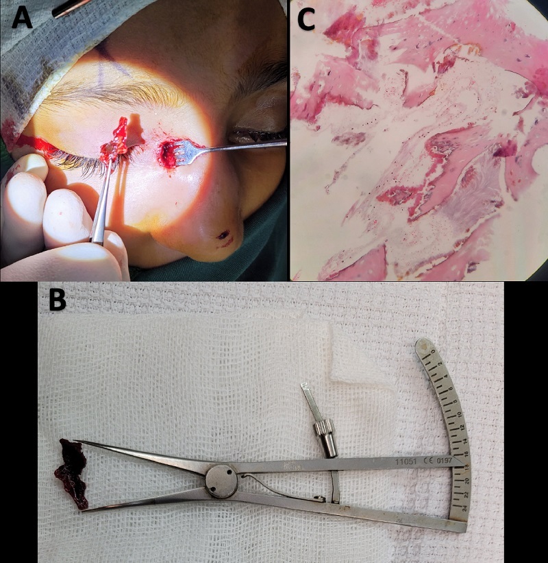
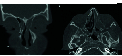
Case report: A nine-year-old girl presented with intermittent unilateral hemolacria and episodes of dacryocystitis with no further remarkable medical or surgical history. CT scan indicated the presence of well-defined calcified tissue enclosed within the lacrimal sac. Following external dacryocystorhinostomy, well-formed firm tissue resembling bony tissue was extracted and sent for pathological evaluation, along with biopsies from the lacrimal sac. Histopathological analysis revealed heterotopic bone formation with nonspecific inflammation of the lacrimal sac. No underlying cause was discernible in the complementary assessment, and no recurrence was noted at one-year follow-up.
Conclusion: A child with hemolacria was found to have heterotopic ossification in the lacrimal sac with no discernible underlying cause.

 求助内容:
求助内容: 应助结果提醒方式:


