{"title":"伪装成黑色素瘤的良性苔藓样角化病的皮肤镜及紫外线反射镜检查。","authors":"Aravind Baskar Murthy, Kallepu Rishika Sagar, Varshini Neelathalli Ramchandraiah, Murali Narasimhan","doi":"10.4103/idoj.idoj_1087_24","DOIUrl":null,"url":null,"abstract":"<p><strong>Abstract: </strong>Benign lichenoid keratosis (BLK), also known as lichen planus-like keratosis, is a benign cutaneous lesion of unknown etiology with overlapping clinical and dermoscopic features with malignant lesions. A 52-year-old female presented with a 10- years history of a single asymptomatic lesion on the left gluteal region not associated with loss of weight, loss of appetite, or other systemic symptoms. Clinical examination revealed a single well-defined violaceous to hyperpigmented plaque of approximately 4 cm × 3 cm in size, on the lateral aspect of the left gluteal region. Dermoscopic examination showed overlapping features of BLK and melanoma. Ultraviolet (UV)dermoscopy showed 'dermoscopic UV blink sign' and areas of blue, yellowish-green, and red fluorescence. Histopathological examination showed features suggestive of BLK with no cellular atypia. The patient was started on 5% imiquimod cream 5 days a week and advised to follow up. This case emphasizes the importance of clinical, dermoscopic, UV reflectance dermoscopic, and histopathological correlation in establishing the diagnosis of BLK masquerading as melanoma with regression, thus aiding in the avoidance of unnecessary surgeries.</p>","PeriodicalId":13335,"journal":{"name":"Indian Dermatology Online Journal","volume":" ","pages":"778-781"},"PeriodicalIF":2.0000,"publicationDate":"2025-09-01","publicationTypes":"Journal Article","fieldsOfStudy":null,"isOpenAccess":false,"openAccessPdf":"https://www.ncbi.nlm.nih.gov/pmc/articles/PMC12419738/pdf/","citationCount":"0","resultStr":"{\"title\":\"Dermoscopy and Ultraviolet Reflectance Dermoscopy of Benign Lichenoid Keratosis Masquerading as Melanoma.\",\"authors\":\"Aravind Baskar Murthy, Kallepu Rishika Sagar, Varshini Neelathalli Ramchandraiah, Murali Narasimhan\",\"doi\":\"10.4103/idoj.idoj_1087_24\",\"DOIUrl\":null,\"url\":null,\"abstract\":\"<p><strong>Abstract: </strong>Benign lichenoid keratosis (BLK), also known as lichen planus-like keratosis, is a benign cutaneous lesion of unknown etiology with overlapping clinical and dermoscopic features with malignant lesions. A 52-year-old female presented with a 10- years history of a single asymptomatic lesion on the left gluteal region not associated with loss of weight, loss of appetite, or other systemic symptoms. Clinical examination revealed a single well-defined violaceous to hyperpigmented plaque of approximately 4 cm × 3 cm in size, on the lateral aspect of the left gluteal region. Dermoscopic examination showed overlapping features of BLK and melanoma. Ultraviolet (UV)dermoscopy showed 'dermoscopic UV blink sign' and areas of blue, yellowish-green, and red fluorescence. Histopathological examination showed features suggestive of BLK with no cellular atypia. The patient was started on 5% imiquimod cream 5 days a week and advised to follow up. This case emphasizes the importance of clinical, dermoscopic, UV reflectance dermoscopic, and histopathological correlation in establishing the diagnosis of BLK masquerading as melanoma with regression, thus aiding in the avoidance of unnecessary surgeries.</p>\",\"PeriodicalId\":13335,\"journal\":{\"name\":\"Indian Dermatology Online Journal\",\"volume\":\" \",\"pages\":\"778-781\"},\"PeriodicalIF\":2.0000,\"publicationDate\":\"2025-09-01\",\"publicationTypes\":\"Journal Article\",\"fieldsOfStudy\":null,\"isOpenAccess\":false,\"openAccessPdf\":\"https://www.ncbi.nlm.nih.gov/pmc/articles/PMC12419738/pdf/\",\"citationCount\":\"0\",\"resultStr\":null,\"platform\":\"Semanticscholar\",\"paperid\":null,\"PeriodicalName\":\"Indian Dermatology Online Journal\",\"FirstCategoryId\":\"1085\",\"ListUrlMain\":\"https://doi.org/10.4103/idoj.idoj_1087_24\",\"RegionNum\":0,\"RegionCategory\":null,\"ArticlePicture\":[],\"TitleCN\":null,\"AbstractTextCN\":null,\"PMCID\":null,\"EPubDate\":\"2025/8/13 0:00:00\",\"PubModel\":\"Epub\",\"JCR\":\"Q3\",\"JCRName\":\"DERMATOLOGY\",\"Score\":null,\"Total\":0}","platform":"Semanticscholar","paperid":null,"PeriodicalName":"Indian Dermatology Online Journal","FirstCategoryId":"1085","ListUrlMain":"https://doi.org/10.4103/idoj.idoj_1087_24","RegionNum":0,"RegionCategory":null,"ArticlePicture":[],"TitleCN":null,"AbstractTextCN":null,"PMCID":null,"EPubDate":"2025/8/13 0:00:00","PubModel":"Epub","JCR":"Q3","JCRName":"DERMATOLOGY","Score":null,"Total":0}
Dermoscopy and Ultraviolet Reflectance Dermoscopy of Benign Lichenoid Keratosis Masquerading as Melanoma.
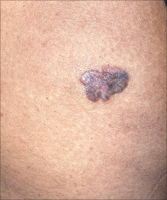
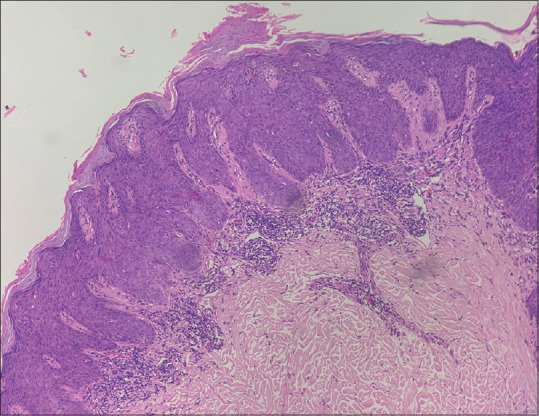
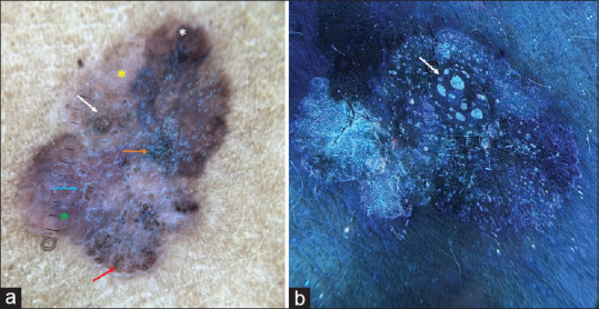
Abstract: Benign lichenoid keratosis (BLK), also known as lichen planus-like keratosis, is a benign cutaneous lesion of unknown etiology with overlapping clinical and dermoscopic features with malignant lesions. A 52-year-old female presented with a 10- years history of a single asymptomatic lesion on the left gluteal region not associated with loss of weight, loss of appetite, or other systemic symptoms. Clinical examination revealed a single well-defined violaceous to hyperpigmented plaque of approximately 4 cm × 3 cm in size, on the lateral aspect of the left gluteal region. Dermoscopic examination showed overlapping features of BLK and melanoma. Ultraviolet (UV)dermoscopy showed 'dermoscopic UV blink sign' and areas of blue, yellowish-green, and red fluorescence. Histopathological examination showed features suggestive of BLK with no cellular atypia. The patient was started on 5% imiquimod cream 5 days a week and advised to follow up. This case emphasizes the importance of clinical, dermoscopic, UV reflectance dermoscopic, and histopathological correlation in establishing the diagnosis of BLK masquerading as melanoma with regression, thus aiding in the avoidance of unnecessary surgeries.

 求助内容:
求助内容: 应助结果提醒方式:


