在唾液腺发育和再生过程中,干细胞/祖细胞动力学被双脉冲追逐范式证明
IF 3.5
3区 环境科学与生态学
Q3 CELL & TISSUE ENGINEERING
引用次数: 0
摘要
活性干细胞和静止干细胞在毛囊和肠隐窝中共存;然而,它们在唾液动力学中的定位和分化潜力尚不清楚。本研究旨在阐明在导管结扎模型中唾液腺发育和再生过程中发生的细胞动力学,重点关注标签保留细胞(lrc),可能是静止干细胞在这些过程中的作用。方法多西环素诱导的tetop -组蛋白2B (H2B)-绿色荧光蛋白(GFP)转基因小鼠[在胚胎第15天(E15) -出生后第7天(P7)诱导GFP表达],然后在P10-14天给药EdU(5-乙基-2′-脱氧尿苷)追踪发育过程中的LRCs。此外,在结扎唾液腺导管前立即用GFP标记lrc,在结扎解除后给予EdU,在组织修复过程中用GFP和EdU追赶lrc。结果和结论在发育过程中,GFP (+) EdU(−)LRCs大量存在于纹状管细胞(SDCs)中,GFP (+) EdU (+) LRCs主要分布于P21的插层管细胞(IDCs)中。标记GFP (+) LRCs和EdU (+) LRCs在P70时褪色。在组织修复过程中,GFP (+) EdU (+) lrc共定位于IDCs和肌上皮细胞(MECs),而随着时间的推移,GFP (+) EdU(+)腺泡细胞(ACs)出现。这些结果表明,唾液腺静止干细胞/祖细胞在发育过程中存在于IDCs中,并且在组织修复过程中,IDCs和MECs中的静止干细胞/祖细胞分化为ACs。本文章由计算机程序翻译,如有差异,请以英文原文为准。
Stem/progenitor cell dynamics during salivary gland development and regeneration demonstrated by the double pulse-chase paradigm
Introduction
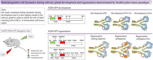
Active and quiescent stem cells coexist in hair follicles and intestinal crypts; however, their localization and differentiation potential in the salivary dynamics are unknown. This study aimed to clarify the cellular dynamics that occur during salivary gland development and regeneration in the duct ligation model with a focus on the role of label retaining cells (LRCs), presumably quiescent stem cells, in these processes.
Methods
Doxycycline-inducible TetOP-histone 2B (H2B)-green fluorescent protein (GFP) transgenic mice [GFP expression was induced during embryonic day 15 (E15)–postnatal day 7 (P7)] followed by EdU (5-ethynyl-2′-deoxyuridine) administration at P10–14 to chase the LRCs during development. In addition, LRCs were labeled with GFP immediately before salivary gland duct ligation and EdU was administered after the ligation was released to chase the LRCs with GFP and EdU during tissue repair.
Results and conclusions
During development, GFP (+) EdU (−) LRCs were abundant in striated duct cells (SDCs) and GFP (+) EdU (+) LRCs were primarily localized to the intercalated duct cells (IDCs) at P21. Labeling of the GFP (+) LRCs faded as well as the EdU (+) LRCs at P70. During tissue repair, GFP (+) EdU (+) LRCs were colocalized in the IDCs and myoepithelium cells (MECs), whereas the GFP (+) EdU (+) acinar cells (ACs) appeared over time. These results suggest that salivary gland quiescent stem/progenitor cells are present in the IDCs during development and that quiescent stem/progenitor cells in the IDCs and MECs differentiate into ACs during tissue repair.
求助全文
通过发布文献求助,成功后即可免费获取论文全文。
去求助
来源期刊
Regenerative Therapy
Engineering-Biomedical Engineering
CiteScore
6.00
自引率
2.30%
发文量
106
审稿时长
49 days
期刊介绍:
Regenerative Therapy is the official peer-reviewed online journal of the Japanese Society for Regenerative Medicine.
Regenerative Therapy is a multidisciplinary journal that publishes original articles and reviews of basic research, clinical translation, industrial development, and regulatory issues focusing on stem cell biology, tissue engineering, and regenerative medicine.
 求助内容:
求助内容: 应助结果提醒方式:


